Day 1 :
Keynote Forum
Joseph Purita
Institute of Regenerative and Molecular Orthopedics, USA
Keynote: Cutting edge concepts in the use of stem cell and PRP injections in an office setting
Time : 09:00-09:35

Biography:
Purita is director of Institute of Regenerative and Molecular Orthopedics (www.stemcellorthopedic.com) in Boca Raton, Florida. The Institute specializes in the use of Stem Cells and Platelet Rich Plasma injections. Dr. Purita is a pioneer in the use of Stem Cells and Platelet Rich Plasma. The Institute has treated some of the most prominent professional athletes from all major sports in both the U.S.A. and abroad. He received a B.S. and MD degree from Georgetown Univ. Dr. Purita is board certified in Orthopedics by ABOS. He is a Fellow American College of Surgeons, Fellow American Academy Orthopedic Surgeons, and a Fellow American Academy of Pain Management. He is also certified in Age Management Medicine. He has lectured and taught extensively throughout the world on the use of Stem Cells and Platelet Rich Plasma. He has been instrumental in helping other countries in the world establish guidelines for the use of Stem Cells in their countries. He has been invited to lecture on these techniques throughout the world as a visiting professor.
Abstract:
The presentation concerns PRP and Stem Cell (both bone marrow and adipose) injections for musculoskeletal conditions in an office setting. Indications are given as to which type of cell and technique to use to accomplish repair. Stem cells, both bone marrow derived (BMAC) and adipose, are used for the more difficult problems. PRP injections are utilized for the less severe problems. Indications are given when to use Stem Cells verses PRP and when to use both. The newest concepts in stem cell science are presented. These concepts include the clinical use of MUSE cells, exosomes, and Blastomere like stem cells. Basic science of both PRP and stem cells are discussed. This presentation defines what constitutes an effective PRP preparation. Myths concerning stem cells are dispelled. One myth is that mesenchymal stem cells are the most important stem cell. This was the initial interpretation of Dr. Arnold Caplan the father of mesenchymal stem cell science. Dr. Caplan now feels that MSCs have an immunomodulation capacity which may have a more profound and immediate effect on joint chemistry and biology. We now learn in the talk that the hematopoietic stem cells are the drivers of tissue regeneration. Also discussed are adjuncts used which enhance the results. These therapies include supplements, LED therapy, lasers, electrical stimulation, and cytokine therapy. The scientific rationale is presented for each of these entities as to how they have a direct on stem cells.
Keynote Forum
Douglas E Garland
Joint Replacement Center, USA
Keynote: In search of excellence: A program, protocols and software for a total joint center with outcomes
Time : 09:35-10:10

Biography:
Garland received his medical degree from Creighton University (1969) and orthopedic surgery residency at Tulane University (1976). He serves on the editorial board of Orthopedics Today, and has been on the clinical faculty at the University of Southern California for over 35 years. Dr. Garland has published more than 100 peer-reviewed scientific articles and chapters. He’s an internationally recognized expert in bone metabolism and his fracture surveys of locations, treatments, and outcomes within orthopedics are considered benchmarks in the field today. Since 2011, Dr. Garland has been the Medical Director for the Joint Replacement Center at Long Beach Memorial.
Abstract:
Background: Joint replacements (JRs) constitute the greatest single cost to Medicare. The majority of JRs in the U.S. are performed by low volume surgeons with a wide variability in clinical and financial outcomes. Joint Replacement Centers of Excellence (JRC) are proliferating, their mission: to provide best/predictable clinical outcomes while reducing cost through efficient/standardized care. It is generally accepted that high volume institutions and surgeons have superior outcomes; however, less is known about the outcomes of participants in JRC. We quantified the effects of surgeon case volume and/or compliance within a JRC program on key hip/knee replacement outcomes with significant financial impact. rnrnMaterials and Methods: During the 2012 calendar year, data of key outcomes for cases performed by two orthopedic surgeon groups performing total hip/or knee replacements at one large community hospital were analyzed: Group I (JRC) includes surgeons who joined the JRC program and Group II (non-JRC) those who did not. Group I was divided into Group IA (JRC >5O) and Group IB (JRC <50) based on surgeons' case volume being more or less than 50 annually. Group I was also divided into Group IC (JRC-Active), comprised of surgeons who regularly attended JRC meetings (>90% attendance) and Group ID (JRC-Passive; <10% attendance). No surgeons in Group II performed more than 50 annual cases. To compare the groups, we chose key outcome variables which have major clinical and financial impacts: blood transfusion rate, discharge to rehabilitation facility versus home, hospital bed days, complications, 30-day readmission, and mortality. rnrnResults: There was a significant decrease in blood transfusion, discharge to rehabilitation facility, and hospital bed days when comparing Group I (N=499) versus Group II (N=96) (p<.001; p<.001; p<.001). Group lA (N=341) versus Group IB (N=158) (p=.001; p<.001; p=.005), Group IC (N=202) versus Group ID (N=297) (p=.007; p=.003; p<.001), and Group IB (N=341) versus Group II (p<.001; p=.004; p<.001). Rates of complications, 30-day readmission, and mortality did not significantly differ among all groups. rnrn Conclusion: Participation in JRC was the major determinant for reduction in blood transfusion, discharge to rehabilitation facility versus home, and hospital bed days. Active/high volume JRC surgeons had the best outcomes. JRC/low volume JRC surgeons far outperformed non-JRC/low volume surgeons. This study is particularly revealing in that low volume surgeons (who perform the majority of joint replacements in the U.S.) can significantly improve certain clinical outcomes and cost savings to the hospital by participating in a well-functioning JRC programrn
- Orthopedic Degenerative Diseases
Arthritis - Types and Treatment
Connective Tissue Disorders and Soft Tissue Rheumatism
Physiotherapy
Session Introduction
Joseph R Purita
Institute of Regenerative and Molecular Orthopedics, USA
Title: The use of supplements and cytokines in platelet rich plasma injections and stem cell treatments
Time : 10:30 -10:55

Biography:
Purita is director of Institute of Regenerative and Molecular Orthopedics (www.stemcellorthopedic.com) in Boca Raton, Florida. The Institute specializes in the use of Stem Cells and Platelet Rich Plasma injections. Dr. Purita is a pioneer in the use of Stem Cells and Platelet Rich Plasma. The Institute has treated some of the most prominent professional athletes from all major sports in both the U.S.A. and abroad. He received a B.S. and MD degree from Georgetown Univ. Dr. Purita is board certified in Orthopedics by ABOS. He is a Fellow American College of Surgeons, Fellow American Academy Orthopedic Surgeons, and a Fellow American Academy of Pain Management. He is also certified in Age Management Medicine. He has lectured and taught extensively throughout the world on the use of Stem Cells and Platelet Rich Plasma. He has been instrumental in helping other countries in the world establish guidelines for the use of Stem Cells in their countries. He has been invited to lecture on these techniques throughout the world as a visiting professor.
Abstract:
The success of stem cell and PRP treatments depends many times on the condition of the extra cellular matrix. The condition of the matrix can have has enormous implications on success or failure on Regenerative Medicine procedures. The matrix is many times a hostile environment of the stem cells. This hostility results in a large percentage of the stem cells (sometimes greater than 97%) perishing. This matrix can many times be manipulated to allow a greater percentage of stem cells surviving. This manipulation can be accomplished by the judicious use of certain supplements, cytokines and other modalities such as micro electrical stimulation and laser use. This talk will center on the use supplements, antioxidants and other modalities. The discussion will also center on free radicals and their mechanisms of causing damage on a cellular level. Also discussed will be certain cytokine pathways which have adirect effect on many orthopedic conditions.
Douglas E Garland
Long Beach Memorial Medical Center, USA
Title: Protocols affect outcomes of both low and high volume surgeons when compared to a control group
Time : 10:55-11:20

Biography:
Garland received his medical degree from Creighton University (1969) and orthopedic surgery residency at Tulane University (1976). He serves on the editorial board of Orthopedics Today, and has been on the clinical faculty at the University of Southern California for over 35 years. Dr. Garland has published more than 100 peer-reviewed scientific articles and chapters. He’s an internationally recognized expert in bone metabolism and his fracture surveys of locations, treatments, and outcomes within orthopedics are considered benchmarks in the field today. Since 2011, Dr. Garland has been the Medical Director for the Joint Replacement Center at Long Beach Memorial.
Abstract:
Background: Patients undergoing joint replacement surgery have higher risk for complications at hospitals with low surgical volume1. Surgeons who perform more than 50 surgeries annually have fewer complications2, 3. The Long Beach Memorial Joint Replacement Center (JRC) is a Destination Center of Superior Performance® created by Marshall Steele/Stryker Performance Solutions® that has a comprehensive course of treatment for persons undergoing elective joint replacement surgery. JRC surgeons have strived for standardization of practice through surgical and post-surgical evidence-based protocols. A retrospective study comparing outcomes of the JRC surgeons and non-JRC surgeons was conducted. Results: In 2012, 11 JRC surgeons performed 584 surgeries compared to 9 non-JRC surgeons who performed 137 surgeries. Four JRC surgeons performed >50 surgeries compared to none of the non-JRC surgeons. A review of specific clinical, operational, and financial outcome measures for elective/non-elective joint replacement surgeries in 2012 demonstrated that there were significant and positive differences between JRC surgeons and non-JRC surgeons in length of stay, discharge home, blood transfusion rates, complication rates, and 30-day readmission rates. Lower direct costs and higher contribution margins were noted for the JRC comparatively. Additionally, JRC surgeons with volumes of less than 50 demonstrated improved clinical outcomes. Conclusion: Strong, collaborative physician leadership in the JRC and establishing evidence-based protocols had positive influences on the clinical outcomes of patients and operational/financial performance of the hospital in JRC surgeons with less than 50 surgeries per year as well as JRC surgeons with more than 50 surgeries per year while non-JRC surgeon outcomes remained unchanged.
Margaret Wislowska
Centralny Szpital Kliniczny MSW, Poland
Title: Antiphospholipid antibody syndrome
Time : 12:10-12:35
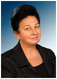
Biography:
Margaret Wisłowska, Head of the Department of Internal Medicine and Rheumatology CSK MSW, the specialist in internal medicine, rheumatology, rehabilitation medicine, hypertention, the author of over 190 scientific papers and books. She has participated in numerous scientific meetings. Promoter of 10 PhD theses. She took trainings at Guy and St Thomas' Hospitals in London, Charity Hospital in Berlin, Rheumatology Institutes in Prague and Moscow. In 2003 she created the Department of Internal Medicine and Rheumatology, and in 2010 Clinic of Internal Medicine and Rheumatology CSK MSW. Prof Margaret Wisłowska is the professor lecturer at the Medical University of Warsaw.
Abstract:
Antiphospholipid syndrome [APS] is the autoimmune disease characterized by vascular thromboses and/or pregnancy loss associated with persistently positive antiphospholipid antibodies (aPL; measured with lupus anticoagulant [LA] test, anticardiolipin antibody [aCL] enzyme-linked immunosorbent assay [ELISA], and/or anti-beta2-glycoprotein-I antibody [alfabeta2GPI] ELISA). Determining significant APS depends on: persistent (at least 12 weeks apart) aPL positivity excluding transient aPL positivity which is common during infections; 2/ a positive LA test is a better predictor of aPL-related thrombotic events compared with other aPL tests; 3/ the specificity of aCL and alfabeta2GPI ELISA tests for aPL-related clinical events increases with higher titers; 4/ 50% of the APS patients with thrombosis present with at least one non-aPL thrombosis risk factor at the time of their vascular event; 5/ IgM isotype is lesse commonly associated with clinical events compared with IgG isotype; 6/ in patients with aPL-related clinical events and no other thrombosis risk factors and have IgAaCL and IgAalfabeta2GPI positivity; 7/triple aPL positivity (LA, aCL, and alfabeta2GPI) can be clinically more significant than double or single aPL positivity. Clinical manifestation related to aPL represent a spectrum: 1/ aPL positivity without clinical events; 2/ aPL positivity solely with non – criteria manifestations (e.g. thrombocytopenia, hemolytic anemia, cardiac valve disease, aPL nephropathy); 3/ APL based on arterial / venous thrombosis and/or pregnancy morbidity; 4/ catastrophic antiphospholipid syndrome.
Eran Maman
Tel Aviv University, Israel
Title: New treatment modality for massive Rotator Cuff tears
Time : 13:25-13:50

Biography:
Eran Maman, Medicine Doctor (M.D), now is a Head of the Shoulder Surgery Unit at Tel Aviv Medical Center. After finishing medical school; Dr.Maman completed his residency in orthopedics. Furthermore, he did a Clinical fellowship in shoulder surgery at Toronto University, Canada. Dr. Maman's research focuses on tendon biology and tendon to bone healing. He aims at finding an optimal biological treatment that is capable of improving tendon-bone interface and promotes healing. He has been working on rat models rotator cuff tears and the influence of many different drugs/material (PRP, steroid, NSAID, statins) on tendon to bone healing in terms of histology and biomechanics. Our group has pioneered on the influence of statins with or without NSAID on the tendon to bone healing on repaired RC.
Abstract:
The treatment of full-thickness massiverotator cuff tears (MRCT) is challenging and associated with a high treatment failure and re-tear rate.As there is no current consensus or definitive guidelines concerning the treatment of this devastating condition, there is a need to evaluate potential alternatives for this patient’s population.The InSpace™ device is a novell treatment modality of an inflatable biodegradable implant, made of a copolymer of Poly Lactic acid and Caprolactonethat degrades within12 months.The spacer is deployed arthroscopically into the sub-acromial space and allows smooth gliding of the humeral head against theacromion. The temporary lowering of the humeral head during spacer inflation in patients with full thickness massive RCTsmay additionally provide improved balance between the subscapularis anteriorly and the infraspinatus posteriorly, permitting better deltoid activation and compensation. The device is approved for use in the EU (since July 2010) and has been tested in several clinical trials as well as implanted in over 4000 commercial cases. The gained clinical experience showed low risk and good safety profile along with a promising effectiveness results, which includesclinically and statistically significant improvementin shoulder functionality, that maintained for a long term (of up to 5 years) in the majority of the treated patients. The use of the InSpace device may be a simple and less invasive alternative that has the potential to provide comparable safety and effectiveness profile to otheravailable surgical options such as arthroscopic partial repair, tendon transfer rotator cuff allograft or arthroplasty.
Rui Shi
Southeast University, China
Title: Characterization and risk factors analysis for reoperation after Microendoscopic Discectomy to treat Lumbar Disc Herniation
Time : 13:50-14:15

Biography:
Rui Shi has completed his MD at the age of 24 years from Southeast University and is now a Ph.D. candidate of Medical School of Southeast University.
Abstract:
A population-based database was analyzed to identify the causes, characteristics of reoperations and associated risk factors after microendoscopic discectomy(MED) to treat lumbar disc herniation(LDH).A series of 952 patients who underwent MED for single-level LDH between 2005 and 2010 were included in this study. Out of this series, 58 patients had revision spinal surgery. The causes and clinical parameters including the intervals between primary and reoperations, grade of disc degeneration, and surgical findings in the revisions were retrospectively assessed. The possible risk factors including age, sex, weight, occupation, duration of surgery, blood loss and radiological findings were evaluated by multivariate logistic regression analysis. In total, 76 disc herniations were excised in revision discectomies with or without interbody fusion for the most common reason-recurrent disc herniation or epidural scar. The overall mean interval between primary and revision surgeries was 39.05 months(range, 2 months to 95 months ). Cumulative overall reoperation rate at 1, 3, 5 years were 1.56%, 2.74%, 5.23% respectively, and gradually increased to 8.17% after near 10 years. Compared to the non-reoperated patients, re-operated patients had older age, higher level of lumbar degeneration, with severe Modic change(Grade â… 17.2%, Grade â…¡ 34.5%, compared with Grade â… 1.5%, Grade â…¡ 30.6% in single-operated patients) and obvious adjacent disc degeneration(81.1%, higher than single-operated patients’ 48.1%). By logistic regression analysis,adjacent segment degeneration and Pfirrmann grading for disc degeneration were identified as significant risk factors related to reoperation after primary MED(OR 2.448, 1.510 respectively).Our study presented a relatively low incidence of reoperation after primary MED. Adjacent segment degeneration, Pfirrmann grading for disc degeneration seem to be the most important risk factors for reoperations after MED to treat LDH. The treatment options for patients with these factors at first visit should be carefully measured.
Neven Mahmoud Taha Fouda
Ain Shams University, Egypt
Title: Obstructive sleep apnea in patients with Rheumatoid Arthritis: Correlation with disease activity and pulmonary function tests
Time : 14:15-14:40

Biography:
Neven Mahmoud Taha Fouda is a Professor of Physical Medicine, Rheumatology and Rehabilitation, Ain Shams University hospitals .She did her M.D degree in November 2003 in Ain Shams University-Cairo-Egypt. She has license to practice medicine in Egypt and worked as consultant of Physical Medicine, Rheumatology and Rehabilitation in many hospitals in Saudi Arabia. She has experience in intra-articular and extra-articular injection and diagnosis by nerve conduction and electromyography.She is also active member in the Egyptian society of Rheumatology and Rehabilitation and Egyptian society of joint disorders and arthritis.
Abstract:
Aim of the work:To assess obstructive sleep apnea (OSA) as one of common primary sleep disorders in patients with rheumatoid arthritis (RA) and its correlation to disease activity and pulmonary function tests. Patients and methods:This study included 30 female patients with RA fulfilled the modified American college of Rheumatology (ACR) criteria.All the patients were subjected to full medical history,thorough clinical examination with evaluation of the disease activity using Disease Activity Score 28(DAS28),laboratory assessment of highly sensitive C reactive protein (hsCRP), pulmonary function tests (FVC- FEV 1 and FEV 1/FVC) and one night polysomnography (PSG) at the sleep laboratory. Results: Polysomnographic data revealed OSA in 14 RA patients ( 46.7%) . Patients with OSA showed longer disease duration (mean 7.0±1.94 y) ,higher BMI (mean 30.8±2.48), higher hsCRP(6.7+0.6 mg/L)and higher DAS28 (4.9 ±1.85) than patients with no OSA (mean 4.0 ±1.72 y, 20.3 ±1.55, 4.9+0.3mg/L and 3.7± 1.28 respectively).While there was statistically non significant difference between both groups as regards results of pulmonary function tests (p>0.05).The study showed statistical significant correlation between AHI (apnea- hypopnea index) and BMI ,hs CRP and DAS 28 (r=0.45 ,0.43 and 0.51 respectively p<0.05), Conclusion: OSA is commonly associated with patients with RA .This possibly suggest common underlying pathological mechanisms which may be linked to chronic inflammation Co-existence of OSA in RA patients will influence the disease activity and the level of circulating inflammatory markers in these patients .Diagnosis and treatment of OSA in RA patients may help in improved clinical care,better prognosis and avoid rheumatoid-associated morbidities.
Shoaib Khan
University Hospital of North Tees, UK
Title: Is cervical plate necessary for anterior cervical fusion? Radiological analysis of fusion rate for anterior cervical discectomy and fusion without plating
Time : 14:40-15:05

Biography:
Shoaib Khan has completed his M.B.B.S from Dow Medical College, Karachi, Pakistan in 2005 and his M.R.C.S from Royal College of Surgeons of Edinburgh, UK in 2013. He is working as a Research Fellow for Spine in University Hospital of North Tees, Stockton on Tees, United Kingdom
Abstract:
Background Anterior cervical discectomy and fusion (ACDF) is considered to be the gold standard treatment for cervical degenerative disease. Different modalities and instrumentation have been used to achieve fusion. The objective of our study was to evaluate the rate of fusion in patients who underwent Anterior Cervical Discectomy and Fusion without the use of cervical plate. Methods and materials The study involved retrospective radiographic analysis of patients who underwent ACDF using cages without plate from August 2005 to February 2014. The radiographs were assessed for fusion independently by a Consultant Radiologist and Consultant Spinal Surgeon using Brantigan-Steffee fusion criteria. The criteria include a denser and more mature bone fusion area than originally achieved at the time of operation, no interspace between the cage and the vertebral body, and mature bony trabeculae bridging the fusion area. The procedures were performed in our unit. Results Thirty nine patients underwent ACDF without plating. Out of 39, 21 were females and 18 were males. Average age for our patients was 62.03 with an average follow up of 25.8 months. Five patients were excluded from study as they had inadequate follow up to comment on fusion. 10 patients had fusion performed at one level, 27 at two levels, one each at 3 and 4 levels. The operated levels for one level patient was C3/4, 4/5, 5/6 and 6/7, for two levels were C3/4 and 4/5, C4/5 and 5/6, C5/6 and 6/7, for three levels was C3/4,4/5,5/6 and for four levels was C3 to C7. Independent analysis by Radiologist and Spinal surgeon showed that fusion was achieved in 28 patients (82%) at all levels, non union was observed in 3 patients(9%), one level (C4/5 and C5/6) was fused out of two levels (C4/5, 5/6 and C5/6, 6/7) in 3 patients(9%). Conclusion ACDF using cages without instrumentation has revealed excellent rate of fusion (82%) which shows that plating is not necessary to attain a better outcome from ACDF.
Sagaram Uday Shanker
Maruthi Rheumatology Research Center, India
Title: Sjogrens syndrome and Hyperlipoproteinemia (a) A detrimental association
Time : 15:50-16:15

Biography:
Uday Shanker received medical degree from Gandhi Medical College, Hyderabad, India in 1978. He then worked as a Sr. Intern in Cardiology in Gandhi Hospital, Hyderabad. In 1984 he worked as a Research associate in Dpt. Nephrology at Thomas Jefferson University Hospital, Philadelphia. He returned to India to do extensive clinical work in Cardiology and Nephrology and started his own Cardiac emergency ICCU where he treated over 10,400 Myocardial Infarctions. He worked in Cardiac Catherization unit, Apollo Hospital, Hyderabad where he produced a paper on Hibernation Myocardium and won fellowship in American College of Angiology. In 2005, he won his first national award, Bharatiya Chikitsak Ratan in New Delhi. Subsequently he won 14 awards including a felicitation and award in London and Bangkok. His passion to serve the rural India made him travel extensively as a consultant Rheumatologist treating around 13000 patients.
Abstract:
INTRODUCTION:
In my 30 years of experience in the field of Cardiology and Rheumatology, I have come across several cases of Dyslipidemia, Hyperlipoprotienemia (a) and inflammatory arthritis. Dyslipidemia not responding to regular treatment with statins, were investigated further and found to have higher levels of lipoprotein (a) which is detrimental to the arthritis patients. On further investigations, few patients were found to have an uncommon combination of Sjogrens syndrome and hyperlipoprotienemia. Such association may lead to sudden / early death. :
OBJECTIVE: Identification of the Association of Hyperlipoprotienemia (a) with Sjogrens syndrome and Vasculitis in autoimmune arthritis diseases.:
METHOD USED: Clinical OP basis: Identified seven cases of Inflammatory arthritis like RA, SLE, MCTD, Enteropathic Arthritis, Psoriatic Arthritis etc. and their association with Hyperlipoprotienemia (a) and associated Sjogrens syndrome ( period 2009 –2015 ):
MEDICAL TREATMENT: - Inflammatory arthritis – DEMARDS and Deflazacort - Hyperlipoprotienemia (a) - Niacin NF 1 gm. per day and Omega fatty acids 500mg per day - Associated dyslipidemias - Statins - Associated diabetes (if required) - Oral Hypoglycemics - Associated hypothyroidism (if required) - Thyroxine tablets
RESULT: Sjogrens syndrome: There was symptomatic relief, such as correction of Dryness of Oral Cavity, Dyspepsia Retroorbital pain, Preauricular Glandular enlargement, lubricant eye drops to dry Palpebrae
- Lipoprotein (a) levels reduced to optimum values in 3 to 6 months:
CONCLUSION:Though very rare, the association of Hyperlipoprotenemia (a) with Sjogrens syndrome and Vasculitis in autoimmune inflammatory arthritis does exist, and the incidence is more in rheumatoid arthritis when compared to SLE, MCTD, Scleroderma and Psoriatic arthritis.
Garima Gupta
Saaii College of Medical Science and Technology, India
Title: Impact of osteoarthritis on balance, perceived fear of fall and quality of life
Time : 16:15-16:40

Biography:
Garima Gupta is result oriented physiotherapist. She is presently working as a Head of Department, Researcher and Assistant Professor in Saaii College of Medical Science and Technology, India. She has done her graduation from super specialty HOSMAT Hospital, Bangalore. In 2010 she completed her Masters in Physiotherapy (Neurology) from Indian Spinal Injury Center, New Delhi. She is actively involved in various ongoing research projects and has multiple international books and research publications in the field of physiotherapy. She actively contributes as reviewer for many international journals. She has also presented two papers in international conferences
Abstract:
Background: Osteoarthritis is the commonest form of joint disease. Reviews suggest presence of balance deficits in osteoarthritic population but in most of the studies expensive force platform, or balance master were used . In the present study we aimed to study the impact of knee OA on balance using cost effective “postural sway-meter”. Present study also aimed to study the coreleation of severity of knee disabilities with perceived fear of fall, previous number of falls and quality of life in OA population. We also aimed to study the affect of balance deficits on quality of life. Methods: 60 people of 50-70 years were taken. Assessment of postural sway was done by sway-meter, quality of life by arthritis impact measurement scale 2 short form, severity of OA by osteoarthritis index of severity and perceived fear of fall by fall efficacy scale was done. Results: The result of the unpaired ‘t’ test analysis showed that people with OA have significant balance deficits when compared to the control group under both eyes open and closed on floor condition. Degrees of knee disability have significant impact on fear of fall and quality of life. Balance deficits have significant impact on patient’s quality of life and their perceived fear of fall. Conclusion: While attending patients with osteoarthritis possible balance deficits should be kept in mind and equal importance should be given to the patient’s fear of fall, quality of life and their severity of knee osteoarthritis
Rutvik Pandya
Rajiv Gandhi University of Health Sciences, India
Title: Global postural re-education
Time : 16:40-17:05

Biography:
Rutvik Pandya is a physical therapy student of Shree Devi College of Physiotherapy, Mangalore, Karnataka affiliated to Rajiv Gandhi University of Health Sciences, Karnataka, India. He has obtained certification for various fitness instructor training like aerobics, spinning, diet and nutrition, primary and advance pilates, pre and post-natal fitness and advance fitness from IAFT- Indian Academy of Fitness Training. He worked as a Personal Physical Trainer for six months at Talwalkars Better Value Fitness Limited, India. He also has attended several workshops related to Physical Rehabilitation, Physical fitness and Awareness.
Abstract:
Global postural re-education (GPR) is a therapeutic method which is exclusively manual and does not require use of machines for the correction and treatment of pathologies in the musculoskeletal system. The aim of this technique is to restore correct alignment of posture and to re-establish a correct biodynamic of body movement in order to prevent or treat musculoskeletal problems. It is based mainly on three methods: Individuality, causuality and globality. The main goal of this technique is to correct the postural deviations. The indications of this technique are any postural deviations, any orthopedic deformities, neurological conditions like dizziness, vertigo, respiratory dysfunctions and post traumatic complications. A GPR session comprises of a series of global stretching positions which evolves gradually from initial position with minimum tension, to a final position with progressive stretching. The frequency and duration of session depends on patient’s problem. Generally it is recommended one session one and half hour per week. This technique is a recent advancement in the field of physical therapy. It is very rarely used in countries like India. It is widely used in countries like Canada, Italy, Spain and Belgium. Although this method is not widely used there are studies done which states that this is a very effective physical therapy method and can be used in the cases like lumbar pain during pregnancy, ankylosing spondylitis, scoliosis, hyperkyphosis, myogenic temporomandibular disorders, stress urinary incontinence, improve cardiovascular responses, chronic neck pain, to improve respiratory muscle strength and thoraco abdominal mobility, idiopathic scoliosis, shoulder protrusion reeducation, stroke, plantar pressure distribution, lengthening the inspiratory muscles, thoracic kyphosis, cervical disc herniation, fibromyalgia.
Gurmeet Singh
Thapar University, India
Title: Investigations for bone surface damage during orthopaedic bone drilling
Time : 17:05-17:30

Biography:
Gurmeet Singh is presently Research Scholar in Mechanical Engineering Departmentat Thapar University, Patiala-Punjab, India. He is working on bone drilling during orthopedic surgery. He has completed his Masters of Engineering in Mechanical Engineering from PEC University of Technology, Chandigarh, India. He has publish 3 international journals and present paper at 4 international conferences. He is having 7 years teaching and research experience. His research area is modern manufacturing, non-conventional machining and bone drilling.
Abstract:
Orthopaedic bone surgery is a curious topic for research in present medical engineering. Machining to bone is very necessary action to treat some major bone fracture. Machining to bone includes through holes, blind holes and sometime just finishing to the bone edges. This machining to the bone can damage the bone and its surroundings if execution is not in a proper manner. This damage may lead to failure of bone joint after some time when human tries to do his daily work, so to maintain the bone joint for long time machining damage should be controlled at the time of machining only. Major problem initiates with machining of bones are crack initiation and thermal damage. This study mainly focuses to maintain the forces exerted and surface damage to the bone during bone drilling with variation of drilling parameters. Using L9 orthogonal array optimized combination of parameters are suggested which gives less damage to the bone surroundings. SEM images of bone drilling surfaces helps to get the micro level information of bone damage.
Gaetano Scuderi
2NYU Medical Center, USA
Title: Improving response to treatment for patients with DDD by the use of molecular markers

Biography:
Gaetano completed his education from University at Buffalo. Previously he worked in Stanford University, UC San Diego and University of Miami. Currently he is working as Orthopedic Surgeon at Palm Beach Spine and Sport, Cytonics Corporation. Jupiter Florida Hospital and Health Care
Abstract:
Background Context: Protein biomarkers associated with lumbar disc disease have been studied as diagnostic indicators and therapeutic targets. A cartilage degradation product, the Fibronectin-Aggrecan complex (FAC) identified in the epidural space, has been shown to predict response to lumbar epidural steroid injection in patients with radiculopathy from herniated nucleus pulposus (HNP) and identified in patients with DDD. A therapeutic agent that prevents the formation of the G3 domain of aggrecan will reduce the fibronectin-aggrecan G3 complex and accordingly may be an efficacious treatment. Since the production of G3 domain of aggrecan is catalyzed by different known classes of proteases, a common inhibitor of all of these proteases could be an ideal therapeutic agent. Such a protease inhibitor is found in plasma and synovial fluid, alpha-2-macroglobulin (A2M). Purpose: Determine the ability of FAC to predict response to biologic therapy with concentrated autologous A2M for patients with LBP from DDD Study Design/Setting: Prospective cohort Patient Sample: 24 patients with LBP pain and MRI positive for DDD Outcome Measures: Oswestry disability index (ODI) and visual analog scores (VAS) were noted at baseline and at 3-month follow-up. Primary outcome of clinical improvement was defined as patients with both a decrease in VAS of at least 3 points and ODI >20 points. Methods: All patients underwent lavage for molecular discography and delayed FAC analysis and injection of platelet poor plasma rich in A2M at the time of the procedure. Results: There were 13 males and 11 females. Age range 24-62 (ave 44.3) 13 pts had 1 level, 6 pts 2 level, and 5, 3 level procedures. 12 discs were FAC + in 10 pts, out of 40 discs tested. 11 pts improved, versus 13 who did not. Patients with FAC-positive assays were significantly more likely to show improvement in their VAS and ODI at follow-up. Mean VAS improvement in FAC-positive patients was 4.9 +/- 0.9, compared to 1.5 +/- 1.2 in those with negative FAC (p < 0.0001; ANOVA). Similarly, ODI improved on average 37 +/- 9.3 points in FACT-positive patients compared to 9.4 +/- 11.9 in FAC-negative patients (p<0.0001; ANOVA). Correlation analysis demonstrated that a FACT-positive test correlates with improvement in VAS (Pearson r = 0.83; p < 0.0001) and ODI (Pearson r = 0.71; p<0.0001). Conclusions: Patients who are “FAC+â€Â are more likely to demonstrate clinical improvement following autologous A2M injection. The results of this investigation suggest that not only is FAC is an important biomarker in identifying who will improve, but also that autologous A2M is an important biologic treatment in discogenic diseases, a true theranostic. We utilized a definition of clinical improvement that was in excess of the minimal clinically improved difference (MCID). Additionally, our defined outcome measure was a combination of two universally accepted outcome parameters (ODI and VAS). The current study bridges the gap between the presence of a biomarker and clinical outcomes.
Jose Miguel Gomez T
University of Minesotta, USA
Title: Comparison of treatment for idiopathic scoliosis based on 2D radiographic analysis and the GOSS system
Time : 11:20-11:45

Biography:
José Miguel Gómez, Profesor, MD, LO. Medical Surgeon from the Pontificia Universidad Javeriana in Bogotá, Colombia. He attended to the Century College, Minnesota for Orthotic program, and completed his orthotic residency at Gillette Children’s Hospital in Saint Paul, Minnesota. Licensed in Orthotic. He has lectured internationally.Founder and currently the Scientific Director of Laboratorio Gilete in Bogotá, Colombia and the President of Gomez Orthotic Systems, LLC Saint Petersburg FL, Assistant Professor, teaching the spinal course at Saint Petersburg College FL, North Western University IL ,South Western University TX, Pittsburg University PA, and Century College MN.Received from American Academy of Orthotics and Prosthetic, the Clinical Creativity Award 2012, in Atlanta
Abstract:
Scoliosis is a spinal curve that has more than ten degrees on the coronal plane, with a rotational component. This means scoliosis is a three dimensional deformity. To start treating a patient, the first thing needed to acknowledge is that the treatment will be done on a person, who is concerned, and is putting their health on the hands of clinicians. The treatment has to incorporate the whole patient, not the spine only. The Gomez Orthotic Spine System is a clinical protocol which measures the patient and treats them with a conservative method used for spinal deformities. The measurements, photos, and x-rays evaluated are used all in analyzing and designing the three dimensional mold. This article will demonstrate the efficacy of the system’s implementation with a single clinical case. It will focus heavily on alignment, balance, and increasing the patients stability by using the center line of each corporal plane
Mithun Neral
Case Medical Center, USA
Title: Silicone arthroplasty of the metacarpo phalangeal joint
Time : 11:45-12:10

Biography:
Mithun Neral is an orthopaedic surgery resident at University Hospitals/Case Medical Center with a particular interest in upper extremity surgery. He attended medical school at the University of Pittsburgh where he studied long-term outcomes of silicone metacarpophalangeal joint arthroplasty.
Abstract:
Abstract Introduction: Silicone arthroplasty of the metacarpophalangeal joint (MCP) is a well-established treatment for rheumatoid arthritis. However, available literature on treatment of non-rheumatic arthritis is limited to case reports and retrospective reviews of small patient populations. Purpose: The purpose of this study is to evaluate the clinical effectiveness of MCP arthroplasty for non-rheumatic arthritis in the largest group of patients with the longest follow-up period to date. We predict that MCP arthroplasty for non-rheumatic arthritis shows significant improvement in hand function, pain relief and patient satisfaction. Materials & Methods: A search of all MCP arthroplasties performed by a single surgeon for non-rheumatic arthritis over a 12-year period found 136 arthroplasties. Of these, adequate prospective follow-up assessment could be completed for 30 patients with 38 MCP arthroplasties at an average of 56 months after surgery. Objective measures included are range of motion (ROM), grip and pinch strength, disabilities of the arm, shoulder, and hand (DASH) score, and visual-analog pain score. Follow-up x-rays were reviewed. Patients also completed a subjective patient-satisfaction questionnaire. Mean ROM, DASH, and pain were compared between the pre-operative and follow-up groups by paired T-test and linear regression to identify significant differences and trends in long-term follow-up. Results: There was significant improvement between mean pre-operative and follow-up ROM, DASH, and pain with p-values of 0.0006, 0.0007 and <0.0001, respectively. Mean follow-up ROM, DASH, and pain scores were 69.5±3.0, 15.0±2.3 and 0.76±0.2, respectively. Linear regression showed significant correlations between pre-operative measurements and improvement at follow-up for ROM, DASH, and pain with p-values of 0.0003, 0.0310, and <0.0001, respectively. No significant difference existed for grip (p=0.593) or pinch (p=0.296) strength when follow-up operative and non-operative hand strengths were compared. The patient-satisfaction questionnaire showed 73% were “very satisfiedâ€, 87% would “definitely do it againâ€, and 70% experience “rare or no pain.†Follow-up x-rays showed 5° mean angulation and 2 mm mean subsidence compared to immediate post-operative X-rays. Four arthroplasties required revision, for an 11% revision rate. Conclusions: This study showed improved ROM and DASH score, excellent pain relief, and excellent patient satisfaction in patients undergoing MCP arthroplasty for non-rheumatic arthritis. Patients with more severe ROM limitation, DASH score and pain score experienced a greater improvement of these measures at follow-up. Strength improvement is limited although remains comparable to the non-operative hand. Angulation, subsidence and complications in the study population are consistent with those reported in current literature.
Ziad Elchami
International Medical Center, Saudi Arabia
Title: The effectiveness of combined radiofrequency (rf) plus epidural therapy versus epidural block alone for spinal stenosis pain due to added hypertrophied lumbosacral facets syndrome

Biography:
Ziad Elchami, Director/Consultant of the Pain and Headache Management Center at the International Medical Center (IMC), Jeddah, KSA since 2005, specializes in chronic pain and neurology; his clinical interests include clinical neurophysiology, neuromuscular disorders, and pain. Dr. Elchami completed his medical degree at the University of Damascus. He then joined Kansas University Neurology residency Program where he completed both his residency and fellowship programs in Clinical Neurophysiology Disorders. During that period, he served as Chief Resident and Chief Fellow. Dr. Elchami has a Pain and Headache Fellowship from the Cleveland Clinic Foundation where he worked from 2003 to 2005.
Abstract:
In lumbar spinal stenosis, spinal nerve roots in the lower back are compressed, producing tingling, weakness or numbness that radiates from the low back, buttocks and legs, while lumbosacral facet syndrome, a type of degenerative arthritis, occurs between the lower back and pelvis, causing significant pain throughout the lower body. The aim of the study is to evaluate the effectiveness of combined radiofrequency (RF) plus epidural therapy versus epidural therapy alone for spinal pain due to added hyperthophied lumbosacral facets syndrome. Study involved 60 patients of Pain & Headache Center, IMC, KSA. First group (N=32) underwent combined LS (RF) + epidural, applied to the lumbosacral facets region, with the following settings: 80 degrees x 1 min and repeated x 3 with repositioning of the needle. Second group (N=28) underwent epidural block only + dexamethasone. Patients were followed up to one year period. Inclusive criteria: 38 females, 22 males; ages between 40-70 years old, with mean of 38 years; and patients who already failed 9-12 sessions of ECSW or PT. Exclusive criteria: patients older than age 80; with uncontrolled diabetes and blood pressure; taking anti-coagulant; other neurological deficits; pregnant women. Average improvement of 75% for the first group, according to the numeric pain scale, was seen in patients who were treated by combined therapy; 60% in second group. Patients with low back pain who went through the combination therapy had more significant improvement than those who went through early epidural block only, with benefits lasting for more than 6 months.
P Karpe
North Tees and Hartlepool NHS Trust,UK
Title: Supramalleolar Osteotomy: A joint-preserving option for advanced ankle osteoarthritis

Biography:
Prasad Karpe has completed his Medical degree from Goa University, India. He then did his Masters of Surgery in Orthopedics from the same university. He has done 4 years of spine fellowships in India and UK. At present, he is doing a Foot and Ankle fellowship as a part of his training. He has also a member of the Royal college of Edinburgh. He has also cleared his Fellowship examinations (FRCS in Trauma and Orthopedics) and is a Fellow of the Royal college of Surgeons of Edinburgh.
Abstract:
Background: Until recently, surgical treatments for advanced ankle osteoarthritis have been limited to arthrodesis or ankle replacement. Supramalleolar osteotomy provides a joint-preserving option for patients with eccentric osteoarthritis of the ankle, particularly those with varus or valgus malaligment. Aim: To evaluate radiological and functional outcomes of patients undergoing shortening supramalleolar osteotomy for eccentric (varus or valgus) osteoarthritis of the ankle. Method: Prospective review of patients from 2008 onwards. Osteotomy was the primary surgical procedure in all patients after failure of non-operative measures. Pre-operative standing antero-posterior and Saltzman view radiographs were taken to evaluate degree of malalignment requiring correction. Radiological and clinical outcomes were assessed at 3, 6 and 12 months post-operatively. Radiographs were reviewed for time to union. Patients were assessed on an outpatient basis for ankle range of motion as well as outcomes using AOFAS scores. Results: 33 patients over a 7 year period. Mean follow-up was 25 months (range 22-30). Mean time to radiological union was 8.6 weeks (range 8-10); there were no cases of non-union. There was a statistically significant improvement in functional scoring (P<0.001); mean AOFAS score improved from 34.8 (range 15-40) pre-operatively to 79.9 (range 74-90) at 12 months post-operatively. There was no significant change in pre- and post-operative range of motion. 2 patients required revision surgery at 12 months; one to arthrodesis and one to ankle replacement. Conclusion: Supramalleolar osteotomy is a viable joint preserving option for patients with eccentric osteoarthritis of the ankle. It preserves motion, redistributes forces away from the affected compartment and corrects malalignment, providing significant symptomatic and functional improvement.
Amir A Beltagi
Cairo University, Egypt
Title: Effect of core stability exercises on trunk muscles time to peak torque in healthy adults
Time : 15:05-15:30

Biography:
Amir A. Beltagi, PT, MSc, is an Assistant Lecturer of Biomechanics, Faculty of Physical Therapy, Cairo University. He is also PhD exchange student at the Biomechanics Research Lab, Department of Orthopedics, College of Medicine, the University of Illinois in Chicago (UIC). His research project is entitled “Biomechanical changes of the spine after induced fixation: a Finite Element". Amir is an active researcher who presented his research work at many international conferences in USA, Germany, Egypt as well as published his research findings in peer-reviewed Journals.
Abstract:
Background: Core stability training has recently attracted attention for optimizing performance and improving muscle recruitment and neuromuscular adaptation for healthy and unhealthy individuals. The purpose of this study was to investigate the effect of beginner’s core stability exercises on the trunk flexors’ and extensors’ time to peak torque. Methods: Thirty five healthy individuals randomly assigned into two groups; experimental (group I) and control (group II). Group I involved 20 participants (10 male & 10 female) with mean ±SD age, weight, and height of 20.7±2.4 years, 66.5±12.1 kg and 166.7±7.8 cm respectively. Group II involved 15 participants (6 male & 9 female) with mean ±SD age, weight, and height of 20.3±0.61 years, 68.57±12.2 kg and 164.28 ±7.59 cm respectively. Data were collected using the Biodex Isokinetic system. The participants were tested twice; before and after a 6-week period during which the experimental group performed a core stability training program. Findings: Statistical analysis using the 2x2 Mixed Design MANOVA revealed that there were no significant differences in the trunk flexors’ and extensors’ time to peak torque between the “pre†and “post†tests for control group (p> 0.05). Also, there were no significant differences in the trunk flexors’ and extensors’ time to peak torque between both groups at the “pre’ test (p>0.05). Meanwhile, the 2x2 Mixed Design MANOVA revealed that there were significant differences in the trunk flexors’ and extensors’ time to peak torque between the “pre†and “post†tests for group I (p<0.0001). Moreover, there were significant differences between both groups for the tested muscles’ time to peak torques at the “post†test (p<0.0001). Interpretation: The improvement in muscle response indicated by the decrease in the trunk flexors’ and extensors’ time to peak torques in the experimental group recommends including core stability training in the exercise programs that aim to improve neuromuscular adaptation and fitness. Keywords: Core stability, Isokinetic, Trunk muscles.
Kun Wang
Southeast University, China
Title: Serum levels of the inflammatory cytokines in patients with lumbar radicular pain due to disc herniation

Biography:
Kun Wang has done his PhD in the Department of Mechanical Engineering at the Tsinghua University, China. He is now an associate professor in the Department of Radiation Oncology at Southeast University ,China.
Abstract:
Purpose: The factors influencing the presence or absence of pain in sciatica secondary to disc herniation remain incompletely understood. We hypothesized that the imbalance in inflammatory cytokines is implicated in the generation of pain. In our study, serum levels of pro-inflammatory and anti-inflammatory cytokines were investigated among patients with severe sciatica; the serum levels were compared with those of patients with mild sciatica and healthy subjects. Methods: In this prospective study, blood protein levels of the pro-inflammatory cytokines, namely, interleukin-6(IL-6), interleukin-8 (IL-8),and tumor necrosis factor-α(TNF-α), and the anti-inflammatory cytokines, namely, interleukin-4 (IL-4) and interleukin-10 (IL-10), of 58 patients with severe sciatica, 50 patients with mild sciatica, and 30 healthy control subjects were analyzed through ELISA. Physical and mental health symptoms were determined using the Oswestry Disability Index (ODI) and Short Form-36 (SF-36) questionnaire. Spearman rank correlation coefficient was also determined to calculate the correlation between the scores obtained from the questionnaires and the serum levels of cytokines. Results: No signiï¬cant difference in IL-6 levels was observed among the three groups; no signiï¬cant difference in IL-8 levels was also found between severe sciatica and mild sciatica groups, although the IL-8 levels of the healthy controls were significantly different from those of the sciatica groups(p<0.001). The TNF-α protein values were approximately fivefold higher in the severe sciatica group than in the mild sciatica group (p<0.01) and the healthy control subjects (p<0.01).The IL-4 protein levels were higher in patients with mild sciatica than in patients with severe sciatica (p= 0.05). The IL-4protein levels were also higher in mild sciatica (p=0.003) and severe sciatica (p= 0.004)groups than in the control subjects. The IL-10 protein values were higher in the severe sciatica group than in the mild sciatica group and the healthy control subjects (p<0.001).ODI was significantly correlated with IL-6 (r= 0.394, p=0.013), TNF-α (r=0.629, p<0.001), and IL-10(r= −0.415, p=0.009). By contrast, ODI was not correlated with IL-4(r= −0.174, p=0.29) and IL-8(r= −0.133, p=0.418). Conclusions: These ï¬ndings support our hypothesis that sciatica pain is accompanied by the imbalance in inflammatory cytokines.
Paul S. Sung
The University of Scranton, USA
Title: A kinematic analysis for shoulder and pelvis coordination in subjects with and without recurrent Low back pain

Biography:
Paul Sung is Associate Professor in Department of Physical Therapy at the University of Scranton, Scranton Pennsylvania. Dr. Sung conducted his research fellowship at the Iowa Spine Research Center, Biomedical Engineering Department at the University of Iowa in Iowa City, Iowa. He is a member of the International Society for the Study of the Lumbar Spine as well as the American Physical Therapy Association. His research interests include the mechanisms of chronic low back pain, sports injury mechanism, spine biomechanics, and non-operative spine care and its clinical application to neuromuscular control.
Abstract:
This presentation is to compare the shoulder and pelvis kinematics based on range of motion (ROM), angular velocity, and relative phase (RP) values during trunk axial rotation. Nineteen subjects with recurrent low back pain (LBP) and 19 age-matched control subjects who are all right limb dominant participated in this study. All participants were asked to perform axial trunk rotation activities at a self-selected speed to the end of maximum range in standing position. The outcome measures included ROM, angular velocity, and RP on the shoulder and pelvis in the transverse plane and were analyzed based on the demographic characteristics between groups. The LBP group demonstrated decreased ROM (p=0.02) and angular velocity (p=0.02) for the pelvis; however, there was no group difference for the shoulder girdle. The ROM difference between the shoulder and pelvic transverse planes had a significant interaction with age (F=14.75, p=0.001). The LBP group demonstrated a higher negative correlation between the shoulder (r=-0.74, p=0.001) and pelvis (r=-0.72, p=0.001) as age increased while no significant correlations were found in the control group. The results of this study indicated that there was a difference in pelvic rotation in the transverse plane between groups during axial trunk rotation. This pattern of trunk movement decreased due to possible pelvic stiffness with neuromuscular constraints. Since subjects with recurrent LBP demonstrated decreased pelvic rotation compared to the shoulder for postural control, increased pelvic flexibility could enhance coordinated movement patterns in order to integrate spinal motion in subjects with recurrent LBP.
Ching-Jen Wang
Kaohsiung Chang Gung Memorial Hospital, Taiwan
Title: Extracorporeal shockwave therapy for Osteoporotic Osteoarthritis of the Knee in Rats. An experiment in animals

Biography:
Ching-Jen Wang, M.D. graduated from National Taiwan University, College of Medicine. He is a board certified orthopedic surgeon and currently holds a clinical faculty at Chang Gung University College of Medicine and serves as a consultant orthopedic surgeon of Kaohsiung Chang Gung Memorial hospital, Taiwan. He has published more than 213 papers in reputed journals and has been serving as the reviewer in many journals. His primary interest and area of expertise include sports medicine, knee and hip replacement surgery, shockwave medicine and tissue regeneration.
Abstract:
The purpose of this study was to investigate the effectiveness of ESWT in osteoporotic osteoarthritis of the knee in rats. Sixty-four female S-D rats were divided into four groups. The control group received sham ovariectomy (OVX) and sham anterior cruciate transection (ACLT) and medial meniscectomy (MM). The osteoarthritis (OA) group received ACLT and MM, but no OVX. The osteoporosis (OP) group underwent bilateral OVX, but no ACLT and MM. The OA + OP group received bilateral OVX, ACLT and MM. One half of animals also received ESWT. The evaluations included the areas of gross pathology, bone mineral density (BMD), micro-CT scan, bone strength test, histopathological examination and immunohistochemical analysis. Group OA + OP showed larger areas of arthritic changes than groups OA and OP as compared to sham group. BMD and bone strength significantly decreased in groups OA, OP and OA+OP versus sham group, and ESWT significantly improved the changes. In micro-CT scan, subchondral plate thickness significantly decreased and bone porosity increased in groups OA, OP and OA+OP, and ESWT significantly improved the changes. .Mankin and Safranin O scores significantly increased in groups OA and OA+OP, relative to sham group, and ESWT significantly improved the changes,. DKK-1 significantly increased, and VEGF, PCNA and BMP-2 decreased in groups OA, OP and OA+OP relative to sham group, and ESWT significantly reversed the changes. This study showed that osteoporosis increased the severity of osteoarthritis of the knee. ESWT was shown effective to ameliorate osteoporotic osteoarthritis of the knee.
Rachel W. Li
Australian National University Medical School, Australia
Title: Heparanase may have a key role in the regulation of inflammatory mediators in Rheumatoid Arthritis

Biography:
Rachel W. Li (MD, PhD) completed her PhD in Australia and gained her postdoctoral experiences in molecular pharmacology focusing on immune regulation of metabolic diseases at the University of Hawaii, the USA. She returned to Australia joining the Trauma and Orthopaedic Research Unit (TORU) at the Australian National University Medical School and has established TORU Laboratory. She is currently leading her team with a focus on osteoimmunology and biomaterials in bone remodeling.
Abstract:
Heparanase is the only known mammalian endoglycosidase capable of degrading the heparan sulfate (HS) glycosaminoglycan, both in extracellular space and within the cell. HS is reported to control inflammatory responses at multiple levels, including the sequestration of cytokines/chemokines in the extracellular space, the modulation of the leukocyte interaction with the endothelium and ECM, and the initiation of innate immune responses. We have reported heparanase expression in synovium of rheumatoid arthritis (RA) patients, and this new finding may offer a new insight of the potential regulatory role of heparanase in the disease activity of RA.However, the precise mode of action by heparanase in inflammatory reactions of RA remains largely unknown. The aim of this project was to examine the heparanase activity, its expression and correlation with the inflammatory mediatory and angiogenic gene expression in plasma and synovium of RA patients, with an ultimate goal of developing heparanase as a potential predictor of RA progression and a new therapeutic target. We have found that a highly significant increase of heparanase activity and expression in synovial fluid and synovial tissue of RA patients, and an increase of the heparanase activity positively correlate with the inflammatory and angiogenic gene expression. We also have some evidence to support a postulation that the involvement of heparanase in gene regulation in the development of pannus in RA may be reflected in a patient’s blood, thus heparanase can be a potential predictor of RA progression and a novel therapeutic target.
Yves Lignereux
Natural History Museum & National Veterinary College, France
Title: Vertebral ankylosing hyperostoses in the horse in archeological contexts: differential diagnosis and comparative nosological review

Biography:
Yves LIGNEREUX, DVM, PhD, Agrege, is Professor of anatomy at the National Veterinary College, Toulouse, France. He has specialized in archeozoology and has been in charge of the scientific programme for the refoundation of the Natural History Museum of Toulouse, reopened to the public in 2008. His current projects in archeozoology are in France, Spain, Cyprus, Egypt, Mongolia.
Abstract:
In the veterinary clinical literature, two basic forms of vertebral fusions have been described: spondyl(arthr)itis/-osisankylopoetica (SPA) and spondylitis/-osischronicadeformans (SPD)(SILBERSIEPE and BERGE, 1958; MORGAN, 1967);advances in clinics have added DISH as a third form, principally in dogs (WOODARDet al., 1985). Inarcheo(zoo)logical contexts, SPA refers to bony bridges connecting synovial joints, while SPD refers to bony bridges between adjacent vertebral bodies(BARTOSIEWICZ, 2013). In human clinics and paleopathology, SPA, SPD and DISH are well-defined entities (e.g. ADLER, 2000; ORTNER, 2003; PAJA, 2012), but with lesional expressions specifically different from animals’. SPA and DISH both are inflammatory, productive, enthesopathic/periosteal connective tissue conditions; SPD is degenerative, erosive, and osteo-/syndesmophytic. SPA is characterized by dorsal and lateral fusions and somatic false-ankyloses; SPD is signaled by spinouspseudarthroses, small-joint osteoarthritis, and disk disease. In all spondyloarthropathies, the ventral longitudinal ligament is highly reactive. Ancient domesticated horses often show lesions of vertebral ankylosing hyperostosis diagnosed as SPA when affecting vertebral bodies and SPD when affecting dorsal arches and their processes. But both processes can interact in a mechanical action-reaction spiral, so that they often coexist on the same vertebra(LEVINEet al., 2005), even questioning the very meaning of differentiating two entities(THILLAUD, pers. com.).Thus, the main supposed etiology is mechanical stress and the causes are looked for in the way horses were handled and utilized. Discussing published cases gives the opportunity to a comparative and critical review of spinal hyperostosing and ankylosing diseases in horse and man.

Biography:
Ole Kudsk Jensen, a specialist of Rheumatology and since 2004 leader of a multidisciplinary research unit of the Spine Center of Silkeborg Regional Hospital, Central Region Denmark. In 2009, he completed his PhD from the University of Aarhus about sensitization of the nociceptive system in low back pain patients. He has published more than 25 papers in international journals, and 12 of these articles have been about sick-listed low back patients. He has been a member of the national Danish board working out guidelines for referral for back surgery.
Abstract:
In 325 sick-listed low back pain (LBP) patients, a multivariate prognostic model for unsuccessful return to work (U-RTW) was developed and validated with satisfying result in a subsequent cohort of 120 patients. These patients were recruited, randomised and managed in the same way as the former group. U-RTW was not different in patients with and without radiculopathy. The model included back+leg pain intensity, side-flexion, bodily distress and four psychosocial risk factors. Magnetic resonance imaging of the lumbar spine was performed consecutively in 141 of these patients. The degenerative manifestations were described standardised, including disc herniations and nerve root compromise. Only type1 Modic changes identified in 18% were negatively associated with U-RTW, also after adjustment for already established risk factors.
Oslei de Matos
Federal Technological University of Parana, Brazil
Title: Influence of sleep disorders on body composition in women with Fibromyalgia

Biography:
Oslei de Matos has completed his PhD in Education and Sports Science at the age of 41 years from Porto University and master degree in Physical Education and Physiotherapy studies from Pontifical Catholic University of Paraná in 1989 and 1995. Medical Gymnastic Specialist in Castelo Branco University of Rio de Janeiro in 1991. He is the director of Laboratory Biochemical and Densitometry, Researcher on women’s health with Osteoporosis, Fibromyalgia and Manager. He is Author of books on postural and physical education. Currently is Anatomy and Biomedical Professor in Graduate Program in Biomedical Engineering in Federal Technological University of Parana in Curitiba – Brazil.
Abstract:
Background: The main objective of this research is relation between sleep disturbance and body composition in women with fibromyalgia. This study used DXA to body composition and Pittsburgh sleep quality index to analyze the sleep quality. This research showed that sample has presence of sleep disturbance with low quality of life, moderate pain. These data showed the relation with excess of fat mass and calories intake. The polysomnography it use to analyze the quality of sleep and to DXA body composition. These data define at what stage of sleep is most affected and the physiological consequences entailed by such phases. Through this analysis correlates with memory loss and alteration of muscle and a greater tendency to fatness components. The main causes of sleep apnea and hypopnea refer to central adiposity, and this will be reviewed after bariatric surgery and improving the quality of sleep after. Other resources used for the diagnosis of fibromyalgia are the Thermography and digital photography. The main objective is the correlation of the pain points with the thermographic analysis of the regions of greatest cellular activity of women with fibromyalgia. The standard are used for the analysis of generalized pain, pain intensity, and quality of life. As the obesity has relation with fibromyalgia, the nutritional assessment it is essential for control of confounding variables in the disease, as well as adaptation to diet for likely improvement of metabolism with increase of antioxidant capacity and facilitation of sleep regulation. All these data are shared with specialized team medical to pharmacological adaptation.
Moin Khan
McMaster University, Canada
Title: Arthroscopic Surgery for Degenerative Tears of the Meniscus: A Meta-Analysis
Biography:
Dr. Khan completed his undergraduate education in the Bachelor of Health Sciences program at McMaster University graduating Summa Cum Laude. He went on to complete his medical training at Michael G. DeGroote School of Medicine in 2010 and graduated from McMaster University's Orthopaedic Surgery Residency Program in 2015. As a recipient of the Margaret and Charles Juravinski Surgical Fellowship award he concurrently completed a Master’s Degree in Health Research Methodology with a specific research focus on clinical outcomes research under the supervision of Dr. Mohit Bhandari and Dr. Olufemi Ayeni. Dr. Khan's research and subspecialty interests relate to orthopaedic sports medicine and in particular the rapidly expanding field of hip preservation surgery. He has published numerous peer-reviewed papers, received national and international media attention for his research, presented at national and international conferences and received multiple research awards for his work.
Abstract:
Introduction: Partial meniscectomy is one of the most commonly performed orthopaedic procedures. Controversy exists with regards to its efficacy in the setting of degenerative meniscal tears. This systematic review and meta-analysis evaluates the efficacy of arthroscopic partial meniscectomy in the setting of mild to no concurrent osteoarthritis of the knee in comparison to non-operative or sham treatments. Methods: Two reviewers independently screened MEDLINE, EMBASE, PUBMED and Cochrane databases for all eligible studies. Only randomized control trials published from 1946 through to Jan 2014 were included. A risk of bias assessment was conducted for all included studies and outcomes were pooled using a random effects model. Outcomes were dichotomized to short-term (< 6 months) and long-term (< 2 years) data. The quality of the evidence was assessed and confidence in recommendations was developed according to the Grading of Recommendations Assessment, Development, and Evaluation (GRADE) approach. Results: Seven randomized control trials were included in this review. Six of the seven included studies had moderate to significant risk of bias. The pooled treatment effect across studies did not demonstrate a statistically significant or minimally patient important difference (MID) between treatment arms for functional outcomes (SMD 0.07, 95% CI -0.10 to 0.23, p=0.43) (I2=20%) or pain relief (MD= -0.06, 95% CI -0.28, 0.15, p=0.57, I2=0%) at two years. Short-term functional outcomes between groups were statistically significant but did not exceed the threshold for MID. (SMD 0.25, 95% CI 0.02 to 0.48, p=0.04). (I2=56%) A priori subgroup analysis related to conservative treatment resulted in substantial decrease in heterogeneity. Sensitivity analyses related to sample size and missing data did not have a significant effect on treatment effect. Conclusions: According to the GRADE approach, there is moderate evidence to suggest there is no benefit of arthroscopic partial meniscectomy for degenerative meniscal tears in comparison to nonoperative or sham treatments for individuals with mild or no concomitant osteoarthritis. A trial of non-operative management should be the first line treatment in such individuals. Future research into identifying indications and ideal patient selection is required with regards to prognostic factors, which may influence outcomes following surgical and conservative treatment.
Abdulkarim Ali
Midland Regional Hospital, UK
Title: Agricultural and Farming Injuries in the Irish Midland
Biography:
Abdulkarim Ali is working in Midland regional hospital, Tullomore, Ireland, UK. He has a interest in Bacterial contamination of Diathermy tips, Intrinsic properties of surgical gloves and Total knee Arthroplasty. He completed his studies from University of Limerick, Ireland, UK
Abstract:
Background: The agricultural and farming business are an important source of employment in the Midlands. This is a retrospective study examining the demographics, characteristics, and outcomes of agricultural and equestrian related injuries presenting to the Midland Regional Hospital, Tullamore, Co. Offaly. Methods: Every presentation to the Accident & Emergency Department at the Midlands Regional Hospital in 2013 was assed retrospectively to determine if an injury had been sustained in an agricultural or equestrian environment. Patient characteristics and injury details were collected for 345 patients who attended the Accident & Emergency Department. Patient demographics, month of occurrence, mechanism of injury, radiology results, management and follow up data were collected and analysed. Results: There were 196 agricultural related presentations to the Accident & Emergency Department and 149 equestrian related presentations. 23% of the agricultural injuries and 36% of the equestrian injuries had confirmed radiological evidence of a fracture or joint dislocation. There were significantly more males involved in agricultural injuries than females (98% vs 2%, p < 0.001). There were significantly more females involved in equestrian injuries than males (58% vs 42%, p < 0.05). 10% of farming injuries and 15.4% of equestrian injuries required admission. Farming machinery accidents contributed to significantly more admissions than any other cause in the agricultural category (p < 0.01). Conclusion: agricultural and farming related injuries in the Irish midland are common presentations to orthopaedic surgeon. Increased attention to occupational health hazards seems required in the equestrian environment as Prevention of adverse health outcomes.
