Day 2 :
Keynote Forum
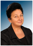
Margaret Wislowska
Centralny Szpital Kliniczny MSW, Poland
Keynote: Rheumatology in the 21st century
Time : 09:00-09:35
Biography:
Margaret Wisłowska, Head of the Department of Internal Medicine and Rheumatology CSK MSW, the specialist in internal medicine, rheumatology, rehabilitation medicine, hypertention, the author of over 190 scientific papers and books. She has participated in numerous scientific meetings. Promoter of 10 PhD theses. She took trainings at Guy and St Thomas' Hospitals in London, Charity Hospital in Berlin, Rheumatology Institutes in Prague and Moscow. In 2003 she created the Department of Internal Medicine and Rheumatology, and in 2010 Clinic of Internal Medicine and Rheumatology CSK MSW. Prof Margaret Wisłowska is the professor lecturer at the Medical University of Warsaw.
Abstract:
Rheumatology in the 21st century specializes in a number of disorders that have defied understanding by the application of current cellular, biochemical and immunologic techniques. The genome revolution does provide us with new tools, which are already beginning to have an impact. Polymorphisms in drugs metabolizing enzymes have been associated with both clinical response and adverse effects of treatment. Proteomics and genomics offer new opportunities to identify biomarkers that provide surrogates of disease activity and response to therapy. MicroRNAs are small, double-stranded RNAs expressed in all cells that regulate the expression of hundreds of genes. MicroRNAs act as master regulators of how a cell responds to changes in its environment, including stresses and inflammation. There have been substantial advances in the field of rheumatology with considerable changes in the management approach of rheumatoid arthritis (RA), spondyloarthritis (SpA), psoriatic arthritis (PsA) and systemic lupus erythematosus (SLE). There is a need to consider the selection of the most appropriate therapy for an individual RA patient and to review how and when to switch treatments in those patients who do not show an optimal response. On 6 November 2012 FDA approve inhibitors of kinases to RA treatment. In SpA the new Assessment of SpondyloArthritis International Society (ASAS) classification criteria have been developed, e.g. classification criteria for axial and for peripheral SpA. In lupus, belimumab, a fully human monoclonal antibody that inhibits B-lymphocyte stimulator BLYSS, was approved for the treatment of lupus in 2011 and there are a number of other therapies in development.
Keynote Forum
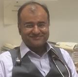
Navtej Sathi
Forth Valley Royal Hospital, UK
Keynote: Is pain crowding the company of orthopaedics and rheumatology?
Time : 09:35-10:10
Biography:
Sathi is currently working as a consultant in Acute Medicine in Scotland. Previously he has previously held posts as a Consultant in Acute Medicine and Rheumatology. His interests include pain, integrated medicine, rheumatology, methotrexate side effects and interstitial lung disease. He holds an M.Sc. in Pain Management from Cardiff University. His research governance score (from Research Gate) is above 20, placing him in the top 30 percent in the world. He is a reviewer for Clinical Rheumatology.
Abstract:
Medical problems related to orthopaedics and rheumatology have been around for thousands of years in the context of humanity. Both specialities have become more defined over the last few centuries. Pain on the other hand has been recognised more as a symptom which has been treated for thousands of years with morphine based drugs as reflected in the traditional World Health Organisation Pain Ladder. For the purpose of the Keynote Speech for Day 2 there will focus on pain and it’s relationship to orthopaedics and rheumatology. Pain is defined by the International Association for the Study of Pain as: An unpleasant sensory and emotional experience associated with actual or potential tissue damage, or described in terms of such damage. The talk will focus on the current published research in the areas pain, distraction and it’s relationship with orthopaedics and rheumatology. Dr. Sathi will talk about his related research work in the field of pain, neuro-oscillations and distraction. A brief review of the modern understanding of pain pathway will also be presented. The idea is to provide a snap shot of the current practice and thinking in this area as well as potentially enthuse the audience to think about ways of reducing pain and thus disability in our orthopaedic and rheumatology patient populations. Thus seeing pain as complementary, and not crowding, to our understanding of patients with orthopaedic and rheumatology problems.
- Ultrasound, Imaging and Diagnostic Techniques
Orthopedic Surgery and Orthopedic Trauma
Musculoskeletal Outcomes: Theory and Practice
Session Introduction
Lloyd S Miller
Johns Hopkins University, USA
Title: In vivo bioluminescence, fluorescence, µCT and photoacoustic imaging tononinvasively evaluate pathogenesis, therapeutics and diagnosticsin an experimentalorthopaedic implant infection
Time : 10:10-10:35

Biography:
Miller completed his M.D. and Ph.D. degrees from SUNY-Downstate Medical Center and his clinical residency and post-doctoral fellowship training at UCLA School of Medicine. He currently is an Associate Professor in the Johns Hopkins Departments of Dermatology, Medicine (Division of Infectious Diseases) and Orthopaedic Surgery. He has published over 30 peer-reviewed manuscripts, including high impact publications in Immunity and Journal of Clinical Investigation. For more than 10 years, Dr. Miller has been a pioneer in utilizing in vivo whole animal optical (bioluminescence and fluorescence imaging), µCT and photoacoustic imaging to study infectious diseases, including orthopaedic implant infections.
Abstract:
Infection isa devastating complication following orthopaedic surgical procedures.Bacteria form biofilmson the implanted materials,creatingchronic infection and inflammation that leads to periprosthetic osteolysis, implant loosening and failure.These infections are challenging to diagnose and treatment is difficult and typically requiresreoperations, prolonged systemic antibiotic courses, extended disability and rehabilitation. To study the these infections, we developed a mouse model of an orthopaedic implant infectionin which a bioluminescent strain of Staphylococcus aureus was inoculated into a post-surgical knee joint of LysEGFP mice (expressing fluorescent neutrophils) that contained a titanium Kirschner-wire extending from the femur. In vivo bioluminescence and fluorescence imaging was used to noninvasivelyquantify the bacterial burden andneutrophil recruitment, respectively. We found that bioluminescent bacteria persisted for the 48 day experiment whereas EGFP-neutrophil signals decreasedwithin the first 14-21 days. Using µCT imaging, the outer bone volume of the femur doubled, which was associated with low-density signals and bone loss, consistent with peri-implant osteolysis.This model was used to evaluate optimal antibiotic combinations to treat an existing infection as well as the novel antimicrobial implant coatings. Finally,as a new diagnostic approach, near-infrared fluorescent probes were used in combination with advanced techniques of photoacoustic imaging. This imaging modalityhas the potential for clinical translation since photoacoustic signals can be detected several centimetersinto tissue. Taken together, bioluminescence, fluorescence, µCT and photoacoustic imaging represent valuable noninvasive techniques to study the pathogenesis of orthopaedic implant infections and to evaluate novel therapeutic and diagnostic strategies.
Artur Yudi Utino
Federal University of Sao Paulo, Brazil
Title: Correlation of the degree of clavicle shortening after non-surgical treatment of midshaft fractures with upper limb function
Time : 10:35-11:00

Biography:
Artur Yudi Utino has completed his medical degree at Universidade Federal de São Paulo, Brazil, in 2011. Concluded his residency in orthopaedics surgery in 2015 and currently is doing a fellowship on Shoulder and Elbow Surgery. Completed his master’s degree in 2015 at the same university
Abstract:
Background: Despite the use of non-surgical methods to treat for the majority of mid-shaft fractures of the clavicle, it is remains controversial whether shortening of this bone following non-surgical treatment of a middle third fracture affects upper limb function. Methods: We conducted a cohort study by sequentially recruiting 59 patients with a fracture of the middle third of the clavicle. All patients were treated non-surgically with a figure-of-eight bandage until clinical and radiological findings indicated healing of the fracture. Functional outcome was assessed using the Disability of Arm, Hand and Shoulder (DASH) score revalidated for the Portuguese language, other outcomes assessed included: pain measured by visual analogue scale (VAS); radiographies to measure the degree of shortening, fracture consolidation and fracture mal-union. Information was also collected regarding the mechanism of injury, patient’s daily activities level and epidemiological features of the patient cohort. The results of our findings are expressed as the comparison of the functional outcome with the degree of shortening. Results: Patients were assessed six weeks and one year after injury. In the first evaluation, the mean DASH score was 28.84 and pain measured by VAS was 2.57. In the second evaluation (one year after injury) the mean DASH score was 8.18 and pain was 0.84. The mean clavicle shortening was 0.92 cm, ranging from 0 to 3 cm (SD=0.64). There was no correlation between the degree of shortening and DASH score after six weeks and one year (p=0.073 and 0.706, respectively). When only patients with of shortening greater than 2 cm were assessed for correlation, the result did not change. Conclusion: We conclude that clavicle shortening after nonsurgical treatment with a figure-of-eight bandage does not affect limb function, even when shortening exceeds 2 cm.
Navtej Sathi
Forth Valley Royal Hospital, UK
Title: Musculoskeletal aspects of hypoadrenalism: Just a load of aches and pain?
Time : 11:20-11:45
Biography:
Sathi is currently working as a consultant in Acute Medicine in Scotland. Previously he has previously held posts as a Consultant in Acute Medicine and Rheumatology. His interests include pain, integrated medicine, rheumatology, methotrexate side effects and interstitial lung disease. He holds an M.Sc. in Pain Management from Cardiff University. His research governance score (from Research Gate) is above 20, placing him in the top 30 percent in the world. His talk on musculoskeletal aspects of hypoadrenalism may provide some insight into how chronic disease can give rise to musculoskeletal symptoms and signs
Abstract:
Hypoadrenalism remains an underdiagnosed disease process. It’s musculoskeletal features are less well described in the literature when compared to diabetes or thyroid disorders. As a consequence considering hypoadrenalism in a patient with mainly musculoskeletal features may be difficult and delayed. The paper will include three cases from our own experience as well as all the relevant cases of hypoadrenalism in the English, French and Spanish language medical literature, going back to the time of Thomas Addison. There will be a summary of the relevant signs, symptoms and investigation findings which may help clinicians diagnose and treat this disease. Also the relationship of hypoadrenalism and chronic disease may be explored during the talk.
Renato A Sernik
Sirio Libanes Hospital, Brazil
Title: Update on the use of ultrasound in the diagnostic of carpal tunnel syndrome
Time : 11:45-12:10

Biography:
Renato A Sernik is a Doctor and obtained his PhD from the University of Sao Paulo. He is a Radiologist at the Medical School Hospital of the University of SaoPaulo for 16 years. He is the author of some books “Ultrasound of the Musculoskeletal System (1999) and Ultrasound of the Musculoskeletal System-Correlation with MRI (2009)”, translated into Spanish and Italian. He is working as a Radiologist at the Sirio Libanes Hospital from 14 years.
Abstract:
Ultrasonography has become one of the main complementary tests for the diagnosis of carpal tunnel syndrome, especially in patients with a history of diseases that may be associated with peripheral neuropathy, such as diabetes mellitus, or in cases where the electroneuromyography is doubtful. The method has also been useful in patients with unsatisfactory results after surgical treatment, where fibrosis, adhesions and neuromas can be identified, having the advantage compared to MRI to be dynamic examination and lower cost. However, the method is operator-dependent with a longer learning curve. Between 2006 and 2014 we analyzed about 120 wrists of patients with clinical suspicion of carpal tunnel syndrome, with electroneuromyographic correlation, being part of the results published in the journal Skeletal Radiology (2008). The increase in cross-sectional area associated with the change in echotexture of the median nerve are the main criteria used for the diagnosis of carpal tunnel syndrome.
Adham Elgeidi
Mansoura University, Egypt
Title: Interleukin-6 and other inflammatory markers in diagnosis of periprosthetic joint infection
Time : 12:10-12:35

Biography:
Adham Elgeidi has completed his MD at the age of 35 years from Mansoura University and postdoctoral studies from both Mansoura University School of Medicine, Egypt and Peninsula University School of Medicine, UK. He is currently a consultant and assistant professor of Orthopaedics and Trauma with special interest in sports medicine and arthroplasty. He is the director of Orthopaedics and Trauma undergraduate teaching and training program Mansoura University. He has published more than 20 papers in reputed journals and has been serving as an editorial board member of repute. He has supervised more than 15 MSc & MD thesis. He has presented more than 30 papers in different conferences. He used to work in Plymouth, UK (May 2003 to August 2006), Tuebingen, Germany (September 2005 to October 2005) and Bordeaux, France (March 2007).
Abstract:
Purpose: to evaluate diagnostic value of interleukin- 6 (IL-6) and other inflammatory markers; C- reactive protein (CRP), Erythrocyte sedimentation rate (ESR), and White blood cell count (WCC) in diagnosis of PJI. Methods: Study group included 40 patients (21 males, 19 females) admitted for surgical intervention after knee or hip arthroplasties. Patients were subjected to careful history taking, thorough clinical examination and preoperative laboratory investigations including serum IL-6, CRP, WCC and ESR. Peri-implant tissue specimens were subjected to microbiological culture and histopathological examination. Results: Mean age of studied patients was 58.4 years (range, 38-72 years). Intraoperative cultures and histopathological examination revealed 11 patients had been infected (PJI) and 29 patients were aseptic failure of prosthesis. Four presumed markers of infection were tested preoperatively: ESR; CRP; WCC; and IL-6. ESR (p=0.0001), CRP (p=0.004), WCC (0.0001), and IL-6 (p=0.0001) were significantly higher in patients with septic revision than those with aseptic failure of the prosthesis. Serum IL-6 (> 10.4pg/ml) reportedly had a sensitivity (100%), a specificity (90.9%), a PPV (79%), a NPV (100%), and accuracy (92.5%). Conclusions: The present study demonstrated that IL-6 has been found to be the most accurate laboratory marker for diagnosing PJI when compared to ESR, CRP, and WCC. IL-6 above 10.4 pg/ml and CRP level above 18 mg/L will identify all patients with PJI and the combination of CRP + IL-6 is an excellent screening test to identify all such patients (sensitivity 100%, NPV 100%).
Tesfaye Hordofa Leta
University of Bergen, Norway
Title: The outcome of unicompartmental knee arthroplasties after aseptic revision into total knee arthroplasties
Time : 12:35-13:00

Biography:
Tesfaye Leta received BEd in Pedagogical Sciencefrom Bahir Dar University, Ethiopia in 1998, BSC inNursing from Bergen University College in 2008, andMPhil in International Health from University of Bergen in 2010. He is working as authorized nurse at the department of orthopeadic surgery, Haukeland University Hospital since 2010: Currently he is a PhD candidate atthe University of Bergen, Bergen,Norway since 2013. The title of his PhD project is Outcome of revision knee arthroplasty in Norway (1994-2011) with special focus on implants survival rate, risk of re-revision, pain, satisfaction, and health related quality of life.
Abstract:
Background: The general recommendation for a failed primary unicompartmental knee arthroplasty (UKA) is revision to a total knee arthroplasty (TKA). Many surgeons prefer to use UKAs in younger patients to postpone TKA believing that the results after revision from a UKA to a TKA is equal to a primary TKA and better than a revision TKA. For this to be true the rev-UKA should outperform the rev-TKA. The purpose of the present study was to compare the outcomes, surgical procedures, and mode of failure of failed primary UKAs and primary TKAs revised to TKAs. Method: The study was based on 768 failed primary TKAs revised to TKAs (rev-TKAs) and 578 failed primary UKAs revised to TKAs (rev-UKAs) and reported to the Norwegian Arthroplasty Register between 1994 and 2011. Patient-reported outcome measures (PROMs) including the EQ-5D, the Knee Injury and Osteoarthritis Outcome Score (KOOS), and Visual Analogue Scales assessing satisfaction and pain were used. Kaplan-Meier and Cox-regression analyses were performed to assess the survival rate and the risk of re-revision. The independent student’s t-test and multiple linear regression were used to estimate the differences in mean scores in PROMs between the two groups. Results: Overall, 12% of rev-UKAs and 13% of rev-TKAs were re-revised between 1994 and 2011. The 10 years survival percentage of rev-UKAs vs rev-TKAs was 82 vs 81%, respectively (p=0.1). There was no difference in the overall risk of re-revision for rev-UKAs vs rev-TKAs (RR= 1.3; p=0.1), nor did we find any differences in the PROM scores. However, the risk of re-revision was 2 times higher for rev-TKA patients aged over 70 years (RR= 2.2; p=0.04). Loose tibia (28 vs 17%), pain alone (22 vs 12%), instability (19 vs 19%), and deep infection (16 vs 31%) were major causes of re-revision for rev-UKAs vs rev-TKAs, respectively but the observed differences were not statistically significant. The surgical procedure for rev-TKAs took longer time (mean=150 vs 114 minutes) and more of the operations needed stems (58 vs 19%), and stabilization (27 vs 9%) compared to rev-UKAs. Conclusion: Despite the surgical procedure of rev-TKA seeming technically more difficult compared to that of rev-UKA, the mode of failure and the overall outcome of rev-UKAs and rev-TKAs were similar. Thus, the argument that the outcome of revising UKA to TKA is similar to primary TKA could not be supported by the present study findings.
Cary Fletcher
St. Ann’s Bay Hospital, Jamaica
Title: Open locked nailing using an expandable nail: An alternative approach
Time : 13:50-14:15

Biography:
Cary Fletcher completed his MBBS degree at University of the West Indies in 2001 and went on to complete his Doctorate in Orthopaedics in 2012, also at The University of the West Indies at the age of 37 years. He is now an Orthopaedic surgeon at St. Ann’s Bay Regional Hospital where he functions as the academic coordinator of the Orthopaedic service. As coordinator, he functions to moderate presentations at all levels of medical staff including residents, interns and student nurses and hence ensure continuing up to date medical education. Doctor Fletcher serves as one of the senior surgeons on the service in which the majority of cases involve Orthopaedic trauma. He is first author on two publications, fifth author on a third and first author on a case report which has been accepted for publication.
Abstract:
Objective: The main objective is to evaluate various outcomes of open intramedullary nailing using the Fixion expanding nail at our institution. Method: A retrospective study was performed using the hospital records. The mechanism of injury, the time between injury and surgery, blood transfusion requirements, blood loss, surgical times, time taken to weight bear (for the femoral/tibial fractures), time for commencement of upper limb use (for humeral fractures), complication rates and the average follow up times were documented. Fifty-seven Long bone fractures in 57 patients were included in this study. Complete results including preoperative X-Rays were available for 27 patients. In 30 cases, the actual X-Rays were not located but documentation by the treating surgeons was available. Results: There were 44 acute femoral fractures, 6 acute tibial fractures, 3 acute humeral fractures, 2 humeral nonunion, 1 tibial nonunion and 1 pathological femoral fracture. All patients achieved radiological union and the complication rates were deemed acceptable. Conclusion: Open intramedullary nailing using an expanding nail may be used for a variety of indications involving the humerus, tibia and femur.
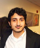
Tariq Alotaibi
King Saud University, Saudi Arabia
Title: Thoracic vertebral hemangioma causing lower limb spastic paresis (case report)
Time : 14:15-14:40
Biography:
Tariq alotaibi : medical student in the final year from king saud university ,riyadh saudi arabia . He is interested in orthopedic. Working on two different research projects in orthopedic.
Abstract:
In this paper we report a rare case of aggressive vertebral hemangioma in an 18-year old male presenting with paraparesis, disturbed sensations and spasticity. Vertebral hemangioma can present with wide variety of symptoms based on extension and compression of the spinal cord. It’s more common in adult. Radiological Imaging were diagnostic of vertebral hemangioma in the level of T8 involving the whole vertebral body. Patient was treated surgically and recovered completely. The clinical presentation, finding on radiological studies, and the management are discussed below.
Abed Al-Negery
Mansoura University, Egypt
Title: Vascularized fibular medialization for reconstruction of the tibial defects following tumor excision
Time : 14:40-15:05

Biography:
Abed Abdel Latif Mohammed Al Negery is a medical Practitioner at Mansoura University hospital and Abed Orthopedic Center, Egypt. He is a professional member of Egyptian Orthopaedic Association and Egyptian Oncology Association.
Abstract:
Purpose: The purpose of this study was to evaluate the functional and oncologic results of fibular medialization when used along as a single-stage reconstructive technique after wide excision of malignant tumors of proximal, middle or distal tibia. Methods: Between December 2010 and May 2015, 14 patients (6 males and 8 females) with primary malignant tumors of the tibia (8 proximal, 4 diaphyseal, 2 distal) were treated by wide excision. The mean age of the patients at the time of surgery was 23.2 years (11 to 38). The fibula was mobilized medially with its vascular pedicle to fill the defect. The average size of the defects reconstructed was 19.5 cm (18 to 22). Full weight bearing was achieved at 6.2 months (range: 4 to 10) postoperatively. Patients were evaluated functionally using the Musculoskeletal Tumor Society evaluation system. Results: The mean follow-up period was 31.3 months (range: 17 to 54). The average time for complete union was 6.2 months (range: 4 to 10). At final follow up all patients had fully united grafts; 9 walked without aids. Chest metastases developed in one patient, superficial wound infection in one patient and leg length discrepancy in three patients. The mean Musculoskeletal Tumor Society Score was 23/30 points (76.5%). The minimum score was 40% and the maximum was 90%. Conclusion: Ipsilateral pedicled vascularized fibular centralization or medialization is a durable reconstruction for tibial defects after wide excision of bone tumors with an acceptable functional outcome. Stable osteo-synthesis is the key to union.
Garima Gupta
Saaii College of Medical Science and Technology, India
Title: Prevalence of musculoskeletal disorders in farmers of Kanpur-rural, India
Time : 15:05-15:30

Biography:
Garima Gupta is result oriented physiotherapist. She is presently working as a Head of Department, Researcher and Assistant Professor in Saaii College of Medical Science and Technology, India. She has done her graduation from super specialty HOSMAT hospital Bangalore. In 2010 she completed her Masters in Physiotherapy (Neurology) from Indian Spinal Injury Center, New Delhi. She is actively involved in various ongoing research projects and has multiple international books and research publication in the field of physiotherapy. She actively contributes as reviewer for many international journals. She has also presented two papers in international conferences
Abstract:
Background: Musculoskeletal Disorders are prevalent and the impact is pervasive across a wide spectrum of occupations, as is evident from numerous studies conducted across the globe. However, there are very few studies that document the prevalence of MSDs in India, and there are hardly any studies that focus on the country’s farming community, which constitutes more than 58 percent of the Indian work force. Thus in the present study an attempt has been made to analyze the prevalence of MSDs in farmers of Kanpur-Rural, India. Methods: A sample of 300 farmers of Kanpur rural district, aged between 20-70 years, was selected. NMQ to measure the musculoskeletal disorders was given to all the farmers. Results: Analysis of data identified four most common MSD affecting the farmers of Kanpur-Rural: LBP (60%); knee pain (39%), shoulder pain (22%), and neck pain (10%). Conclusion: Finding of the present study shows that yearly prevalence of MSDs in farmers of Kanpur-Rural, India is alarmingly high and it suggests that nearly 60 percent of Indian cultivators could be afflicted by this disease, which urgently needs to be corroborated by similar studies at the national level. LBP is the most prevalent type of MSDs affecting the famers. Knee, shoulder and neck pain are other important MSDs affecting farmers in the study area.Observations made during the present study suggest that poor postures and lack of ergonomic awareness in the farming community are the two principal causative factors contributing to the development of MSDs.
Sameer M Naranje
Helena Regional Medical Center, USA
Title: An update on lower limb joint replacement in rheumatoid arthritis

Biography:
Sameer Naranje has completed his fellowship in joint replacement and adult reconstructive surgery from University of Minnesota after completing his M.R.C.S from Royal College of Physicians and Surgeons, Glasgow and Orthopaedic Residency from prestigious All India Institute of Medical Sciences, New Delhi, India. He is currently Chief of Surgery and Orthopaedics at Helena Regional Medical Center, AR, USA and Consultant Orthopaedic Surgeon at East Arkansas Orthopaedic Associates, AR, USA. He has extensive research background, numerous conference presentations, has published more than 30 papers in reputed journals and has been serving as an editorial board member and reviewer of number of reputed Orthopaedic journals.
Abstract:
More than 2/3rd of patients of Rheumatoid arthritis (RA) become disabled 20 years from primary diagnosis. RA is one of the most common indications for lower limb joint replacement in Northern Europe and North America. Though improved medical treatment of RA over last 2 decades have decreased the rate of hip and knee surgery, over a third of patients will need a major joint replacement, of which the majority will receive a total hip or knee replacement (THR and TKR). This paper summarizes an update on the major lower limb arthroplasty surgery for patients with RA. A multidisciplinary approach is needed for preoperative optimization. RA patients may need joint replacement at relatively younger age when compared to the patients with osteoarthritis and may need mutilple revision surgeries over their lifespan. Patients should be made aware of this and increased risk of infection and periprosthetic fracture rates associated with their disease. Biologic agents should be stopped pre-operatively due the increased infection rate. However, continued use of methotrexate does not increase infection risk, and may infact be helpful in recovery. The surgical sequence is commonly hip, knee and then ankle. Cemented total hip replacement (THR) and total knee replacement (TKR) have superior survival rates over uncemented components. Achieving ligamentous balance may be challenging in knee replacements in these patients and more constrained implants may be needed in patients with poor ligaments and severe deformities. The evidence is not clear regarding a cruciate sacrificing versus retaining in TKR, but a cruciate sacrificing component limits the risk early instability and potential revision. The results of total ankle replacement remain inferior to THR and TKR though the science of ankle replacement continues to evolve. RA patients achieve equivalent pain relief after joint replacement, but their rehabilitation is slower and their functional outcome may not be optimum due to continued presence or worsening of the disease. Again, the key to managing these complicated patients is to work as part of a multidisciplinary team to optimize their outcome
Andrew Clerk
Ayr University Hospital, UK
Title: A series of nerve repairs on traumatic nerve injuries
Biography:
I am Andrew Clerk and I work as a Speciality Trainee in orthopaedics. I have been working in orthopaedics for more than 5 years. My qualifications are MB ChB and MRCS.
Abstract:
Nerve injuries are a difficult surgical problem to tackle with. They have been accepted as a entity with poor prognosis. We share our experience of surgical repair of nerve injuries in three different hospitals of South Asia and Western Europe. The objectives of this retrospective study was to audit the results of nerve repairs done within a period of 10 years in three different centres of the world and assess the outcome. A retrospective analysis of the nerve repairs of the upper limb as well as of the lower limb of 456 patients were under taken in this study. The study period was 10 years from January 2003 to October 2013 and involved nerve repairs done in three institutions. Case notes of nerve repairs identified from theatre registers were used to gain information of the relevant patients. Pre-operative assessment notes and operative notes were used to identify the nerves involved, the mechanism of injury and operative interventions undertaken. Follow-up details were obtained also from the records in the case notes of the patients. A total of 456 patients were looked at in three different hospitals in Colombo, Manchester and Ayr. Out of the 456 patients 382 were males and nearly 68% were due to violence. About 12% were due to domestic accidents and 20% were occupational injuries. Out of the nerve injuries 76% were involving the upper limb and a total of 79% were associated with fractures. Three hundred and twenty one patients had primary nerve repairs. Out of the patients who had primary nerve repairs nearly 62% achieved a good functional out come. Of the group of patients who had delayed nerve repairs due to various reasons only 23% achieved a good functional out come. Common causes for nerve injuries are related to violence and occupational hazards. They are more common in the upper limb. Nerve injuries are best dealt with in the primary wound exploration where ever possible for best functional out come. Delayed exploration and repair is not entirely without a good functional result.
Thisara C Weerasuriya
Tameside General Hospital, UK
Title: Sub-laminar wiring in spinal fractures – A viable alternative

Biography:
I have gained experience in general orthopaedics, in trauma as well as in elective orthopaedic practice both in Sri Lanka and in the United Kingdom. My practice in orthopaedics spans nearly ten years from the year 2000 to date of which the majority of my experience was gained in my native country, Sri Lanka. Inaddition to primary and revision joint replacements I am specially interested in instrumentation of traumatic spinal fractures and limb salvage surgery. Specialties:My special interests are Limb salvage and trauma. In the field of limb salvage surgery I have expeprience in salvage and compartmentectomy of limbs involved in bone tumours with allograft recontruction. In the field of trauma my special interest is on salvaging limbs deamed beyond salvage by most salvage scoring systems with emphasis on re-implantation of severed limbs due to traumatic amputations. My current series is 36 limb re-implantations during the past 10 years.
Abstract:
Introduction In the coastal regions of the island nation of Sri Lanka (Ceylon) toddy tapping form coconut trees is a common occupation which is part of the vast industry of production of juggery, coconut vinegar, toddy and arrack( a whisky brewed from coconut sap). The harvesting of the sap is done by toddy tappers who climb these nearly forty foot high palm trees and cross from one tree to another using rope bridges. Falls from these rope bridges are not uncommon as rats that are often attracted to the coconut sap take the same root up the tree and damage the rope bridges that are used by the toddy tappers. Hence falls from these tall trees with resultant spinal fractures is common in the coastal belt of Sri Lanka. I believe that it is important to give a detailed demographic out line of this rather unique problem related to a particular occupation in Sri Lanka. Therefore I think this section should be retained for the information of the readers. Background In Sri Lanka toddy tapping is a common form industry in the coast line villages. Falls from palm trees of forty feet is a common occupational hazard. Spinal fractures are common in these patients . Methods A retrospective analysis of a personal case series of the two authors was undertaken. Results A total of 37 pedicle screw fixations and 58 sub laminar wirings were done during a period of five years from 2004-2009. Follow up time was 6 months to 4 years (mean 3 years). Of the 58 sub laminar wirings, 41 were for lumbar fractures and 17 for thoracic fractures. Thirty two of the 58 sub laminar wirings were for injury due to falls from scaffoldings. Twenty one were toddy tappers who fell off the coconut palms. Five of the 37 pedicle screw fixations were done for thoracic fractures and 32 were done for lumbar fractures. Fourteen patients had falls from scaffoldings. Thirteen posterior fusions of the cervical spine were done. Seven of these were caused by tree climbing accidents. Three patients with sub-laminar wiring developed superficial wound infections while two had to have their metal work taken out due to deep infection. One thoracic sub laminar wiring patient had a CSF leak and two patients with lumbar pedicle screw fixation had cerebro-spinal fluid (CSF) leaks. Three patients with pedicle screw fixations to the lumbar spine had metal work failure due to non-union. One patient with pedicle screws had a re-fracture while three patients with sub laminar wiring of the thoracic spine had pneumothoraxes. Conclusion Sub laminar wiring is a cheap stable method of fixation for traumatic spines which can be used in relatively under- equipped centers as well.
Paul S. Sung
The University of Scranton, USA
Title: Outcome measurement based on kinematic characteristic in patients with low back pain

Biography:
Paul Sung is Associate Professor in Department of Physical Therapy at the University of Scranton, Scranton Pennsylvania. Dr. Sung conducted his research fellowship at the Iowa Spine Research Center, Biomedical Engineering Department at the University of Iowa in Iowa City, Iowa. He is a member of the International Society for the Study of the Lumbar Spine as well as the American Physical Therapy Association. His research interests include the mechanisms of chronic low back pain, sports injury mechanism, spine biomechanics, and non-operative spine care and its clinical application to neuromuscular control.
Abstract:
This presentation is to introduce evidence based kinematic changes in the lumbar spine in subjects with and without recurrent low back pain (LBP) while standing on one leg with and without visual feedback. The lumbar stability index includes relative holding time (RHT) and relative standstill time (RST). Even though a number of studies have evaluated postural adjustments based on kinematic changes in subjects with LBP, lumbar spine stability has not been examined for abnormal patterns of postural responses with visual feedback. The stability index of the core spine significantly decreased in both RHT and RST, especially when visual feedback was blocked for subjects with LBP. The interaction between visual feedback and trunk rotation indicated that core spine stability is critical in coordinating balance control. A trunk muscle imbalance may contribute to unbalanced postural activity, which could prompt a decreased, uncoordinated bracing effect in subjects with LBP. As a result, kinematic rehabilitation training could be used in the prevention of postural instability. The effect of visual feedback on kinematic changes, such as RHT and RST, has not been carefully considered in subjects with LBP. Statistically significant and clinically relevant differences in postural stability and visual feedback were observed between subjects with and without LBP during the one leg standing test. Subjects with LBP have decreased RHT and RST when visual feedback is blocked. As a result, the early detection of kinematic imbalance might be required to understand compensatory mechanisms and postural adjustments in subjects with LBP.
Rajiv Limaye
University Hospital of North Tees and Hartlepool Stockton on Tees, UK
Title: Review of surgical excision of Morton’s inter-metatarsal neuroma using plantar approach

Biography:
He has been in post since January 2006 as Consultant Orthopaedic and Trauma Surgeon at Friarage Hospital Northallerton, a part of South Tees University Hospital NHS Trust. He is a medically qualified doctor, Consultant and a specialist Foot and Ankle Surgeon. He provides a tertiary foot and ankle service at Friarage Hospital, Northallerton. He is on the specialist register of the GMC. Mr Limaye is an Orthopaedic Expert with extensive clinical experience. Due to several requests from solicitors undertaking personal injury work, Mr Limaye started his medico-legal practice in 2005 to provide independent medico-legal reports to solicitors and medical reporting organisations. He is registered as a first-tier medical expert with the Association of Personal Injury Lawyers (APIL) and he is a member of the Leeds and West Riding Medico-legal Society. He has completed hundreds of reports to date.
Abstract:
Introduction Pain in the inter-metatarsal region in the foot due to Morton’s neuroma is a very common presentation in both orthopaedic and rheumatology clinics. Various techniques have been described in the literature for the treatment of this very common condition, including surgery. There is also variation amongst surgeons using various surgical approaches. There are benefits and disadvantages of various approaches. We aimed at reviewing the efficiency of neurectomy by using a plantar approach. 20 patients were surgically treated using this approach. Patients and Methods A series of 20 feet in 20 patients were included in this study. These patients had confirmed Morton’s neuroma in one or two interspaces in their feet. The diagnosis was done by clinical examination followed by an ultra sound examination. In addition, these patients had either an orthotic support or an inter-metatarsal injection as a prior method of treatment. The rest of their feet examination was found to be normal and there were no other causes found to be contributory for their forefoot pain. All of these patients were surgically treated using a plantar incision (either vertical or horizontal) and a neurectomy was performed. The mean age was 47 years. There were 14 females and 6 males. Out of our cohort, 60% of the cases had two level involvement (second/third interspace and third/fourth interspace) and 40% had only one space involvement (second and third interspace.) The average time to resolve the symptoms was 4.5 weeks. The mean follow-up was 8 months. The results were analysed by an independent surgeon clinically using AOFAS scores. The VAS and subjective outcome score were also performed. Results The mean AOFAS score improved from 39 pre-op (median 37, range 20 - 70) to 80 post op. (median 77, range 70 - 93). The mean VAS improved from 40 to 92. Out of the total patients, 90% were very satisfied/ satisfied. The time for resuming normal activities was found to be 4.5 weeks in 80% of patients. There were no cases of infection, residual pain beneath the scar and wound complications. Two patients had no benefit of the procedure and these were rescanned to show evidence of post-operative scarring and bursitis and no further surgery was offered to these patients. There was no evidence of recurrence of neuroma in these patients. The remaining patients were happy at the time of discharge. Conclusion This small series represents the author’s preferred approach towards this common problem in orthopaedics and rheumatology. The authors conclude that neurectomy performed by a plantar approach is satisfactory, because of better neuroma exposure, satisfactory soft tissue healing, faster return to normal activities and pain improvement post operatively.
Piseth Seng
La Conception University Hospital, France
Title: Osteoarticular infection caused by Staphylococcus Lugdunensis: A multi-center study

Biography:
Piseth Seng has completed his Medical Doctor at the age of 24 years from University of Health Sciences in Kingdom of Cambodia, his Specialist in Internal Medicine andhisSpecialists in Tropical Medicine and Infectious Disease at 32 years from Aix Marseille University in France and his PhD at the age of 36 years from Aix Marseille University, France.He is a Physician, Infectious Diseases Unit, La Conception University Hospital, Marseille, France.He has published 14 papers ininternational journals and has been serving as an invited reviewers in international journals and an editorial board member of repute.
Abstract:
Staphylococcus lugdunensis is a virulent coagulase-negative staphylococcus that behaves as S. aureus. S. lugdunensisosteoarticular infection were rarely reported.We report a series of the 119 cases of S. lugdunensisosteoarticular infection managed in 9 hospital centers and 3 private clinics in the South and the Southeastern of France. The median age of patients was 61 years with male-to-female sex ratio at 2.4. We have identified 94 cases (79%) of osteoarticular infectionassociated with orthopedic device including 2 cases of infection after anterior cruciate ligament reconstruction, 62 cases of prosthetic joint infections, 32 cases of internal orthopedic device infection and 3 cases of vertebral orthopedic device infection. Nine-percent of orthopedic device infection were occurred before the1st month afterthe implantation, 15% of cases between 1st-6th months and 52% of cases after the 6thmonths.We have identified 25 cases (21%) of bone and joint infection without orthopedic device including 4 cases of arthritis, 19 cases of osteitis and 2 cases of vertebral osteomyelitis without orthopedic device.Seventy-six percent of cases were treated by surgery including 39% of cases by lavage and debridement, 24% by orthopedic device removal and 17% prosthesis removal. Amputation has been observed in 8% of cases. The remission was observed in 67% of cases; whereas the relapses were observed in 14% of cases. S. lugdunensisosteoarticular infectionis an underestimated hospital-acquired infection which are often associated with orthopedic devices. The orthopedic devices must be removed to control infections embedded in biofilm.
Biography:
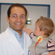
Mohamed M H El-Sayed is a Professor & Consultant of Pediatric Orthopedics & Limb Reconstructive Surgeries in the Faculty of Medicine, Tanta University. He is also a Fellow of LMU - Deutschland and a Fellow of UNT - USA, Member of EPOS, ASAMI, AAOS, IFPOS, EOA.
Abstract:
Background: Evaluation and treatment of the musculoskeletal problems in MMC patients can be quite difficult due to the loss of sensation affecting some or all parts of the lower extremities, associated congenital anomalies of the spine and lower extremities, and muscle imbalance that affects skeletal development over the entire period of growth. Furthermore, some patients who have MMC, have a static encephalopathy that impairs coordination and results in the loss of strength of the lower and upper extremities. In addition, progressive neurological deterioration may occur because of tethered cord syndrome or syringomyelia. Reduction of a dislocated hip in MMC patients was always controversial. Patients & Methods: In this study we present the reduction of 12 dislocated hips in 6 lower lumbar MMC patients, whom we followed for a minimum of 2 years,( 2-7 years). Results: All the patients achieved assisted ambulation within the follow-up period. Significance: MMC patients with lower lumbar lesions (quadriceps function ≥ grade3), are potential community ambulators and this warrants hip reduction and reconstruction in dislocated cases.
