Day :
- Orthopedic Degenerative Diseases
Arthritis - Types and Treatment
Connective Tissue Disorders and Soft Tissue Rheumatism
Physiotherapy
Session Introduction
Joseph R Purita
Institute of Regenerative and Molecular Orthopedics, USA
Title: The use of supplements and cytokines in platelet rich plasma injections and stem cell treatments
Time : 10:30 -10:55

Biography:
Purita is director of Institute of Regenerative and Molecular Orthopedics (www.stemcellorthopedic.com) in Boca Raton, Florida. The Institute specializes in the use of Stem Cells and Platelet Rich Plasma injections. Dr. Purita is a pioneer in the use of Stem Cells and Platelet Rich Plasma. The Institute has treated some of the most prominent professional athletes from all major sports in both the U.S.A. and abroad. He received a B.S. and MD degree from Georgetown Univ. Dr. Purita is board certified in Orthopedics by ABOS. He is a Fellow American College of Surgeons, Fellow American Academy Orthopedic Surgeons, and a Fellow American Academy of Pain Management. He is also certified in Age Management Medicine. He has lectured and taught extensively throughout the world on the use of Stem Cells and Platelet Rich Plasma. He has been instrumental in helping other countries in the world establish guidelines for the use of Stem Cells in their countries. He has been invited to lecture on these techniques throughout the world as a visiting professor.
Abstract:
The success of stem cell and PRP treatments depends many times on the condition of the extra cellular matrix. The condition of the matrix can have has enormous implications on success or failure on Regenerative Medicine procedures. The matrix is many times a hostile environment of the stem cells. This hostility results in a large percentage of the stem cells (sometimes greater than 97%) perishing. This matrix can many times be manipulated to allow a greater percentage of stem cells surviving. This manipulation can be accomplished by the judicious use of certain supplements, cytokines and other modalities such as micro electrical stimulation and laser use. This talk will center on the use supplements, antioxidants and other modalities. The discussion will also center on free radicals and their mechanisms of causing damage on a cellular level. Also discussed will be certain cytokine pathways which have adirect effect on many orthopedic conditions.
Douglas E Garland
Long Beach Memorial Medical Center, USA
Title: Protocols affect outcomes of both low and high volume surgeons when compared to a control group
Time : 10:55-11:20

Biography:
Garland received his medical degree from Creighton University (1969) and orthopedic surgery residency at Tulane University (1976). He serves on the editorial board of Orthopedics Today, and has been on the clinical faculty at the University of Southern California for over 35 years. Dr. Garland has published more than 100 peer-reviewed scientific articles and chapters. He’s an internationally recognized expert in bone metabolism and his fracture surveys of locations, treatments, and outcomes within orthopedics are considered benchmarks in the field today. Since 2011, Dr. Garland has been the Medical Director for the Joint Replacement Center at Long Beach Memorial.
Abstract:
Background: Patients undergoing joint replacement surgery have higher risk for complications at hospitals with low surgical volume1. Surgeons who perform more than 50 surgeries annually have fewer complications2, 3. The Long Beach Memorial Joint Replacement Center (JRC) is a Destination Center of Superior Performance® created by Marshall Steele/Stryker Performance Solutions® that has a comprehensive course of treatment for persons undergoing elective joint replacement surgery. JRC surgeons have strived for standardization of practice through surgical and post-surgical evidence-based protocols. A retrospective study comparing outcomes of the JRC surgeons and non-JRC surgeons was conducted. Results: In 2012, 11 JRC surgeons performed 584 surgeries compared to 9 non-JRC surgeons who performed 137 surgeries. Four JRC surgeons performed >50 surgeries compared to none of the non-JRC surgeons. A review of specific clinical, operational, and financial outcome measures for elective/non-elective joint replacement surgeries in 2012 demonstrated that there were significant and positive differences between JRC surgeons and non-JRC surgeons in length of stay, discharge home, blood transfusion rates, complication rates, and 30-day readmission rates. Lower direct costs and higher contribution margins were noted for the JRC comparatively. Additionally, JRC surgeons with volumes of less than 50 demonstrated improved clinical outcomes. Conclusion: Strong, collaborative physician leadership in the JRC and establishing evidence-based protocols had positive influences on the clinical outcomes of patients and operational/financial performance of the hospital in JRC surgeons with less than 50 surgeries per year as well as JRC surgeons with more than 50 surgeries per year while non-JRC surgeon outcomes remained unchanged.
Margaret Wislowska
Centralny Szpital Kliniczny MSW, Poland
Title: Antiphospholipid antibody syndrome
Time : 12:10-12:35

Biography:
Margaret Wisłowska, Head of the Department of Internal Medicine and Rheumatology CSK MSW, the specialist in internal medicine, rheumatology, rehabilitation medicine, hypertention, the author of over 190 scientific papers and books. She has participated in numerous scientific meetings. Promoter of 10 PhD theses. She took trainings at Guy and St Thomas' Hospitals in London, Charity Hospital in Berlin, Rheumatology Institutes in Prague and Moscow. In 2003 she created the Department of Internal Medicine and Rheumatology, and in 2010 Clinic of Internal Medicine and Rheumatology CSK MSW. Prof Margaret Wisłowska is the professor lecturer at the Medical University of Warsaw.
Abstract:
Antiphospholipid syndrome [APS] is the autoimmune disease characterized by vascular thromboses and/or pregnancy loss associated with persistently positive antiphospholipid antibodies (aPL; measured with lupus anticoagulant [LA] test, anticardiolipin antibody [aCL] enzyme-linked immunosorbent assay [ELISA], and/or anti-beta2-glycoprotein-I antibody [alfabeta2GPI] ELISA). Determining significant APS depends on: persistent (at least 12 weeks apart) aPL positivity excluding transient aPL positivity which is common during infections; 2/ a positive LA test is a better predictor of aPL-related thrombotic events compared with other aPL tests; 3/ the specificity of aCL and alfabeta2GPI ELISA tests for aPL-related clinical events increases with higher titers; 4/ 50% of the APS patients with thrombosis present with at least one non-aPL thrombosis risk factor at the time of their vascular event; 5/ IgM isotype is lesse commonly associated with clinical events compared with IgG isotype; 6/ in patients with aPL-related clinical events and no other thrombosis risk factors and have IgAaCL and IgAalfabeta2GPI positivity; 7/triple aPL positivity (LA, aCL, and alfabeta2GPI) can be clinically more significant than double or single aPL positivity. Clinical manifestation related to aPL represent a spectrum: 1/ aPL positivity without clinical events; 2/ aPL positivity solely with non – criteria manifestations (e.g. thrombocytopenia, hemolytic anemia, cardiac valve disease, aPL nephropathy); 3/ APL based on arterial / venous thrombosis and/or pregnancy morbidity; 4/ catastrophic antiphospholipid syndrome.
Eran Maman
Tel Aviv University, Israel
Title: New treatment modality for massive Rotator Cuff tears
Time : 13:25-13:50

Biography:
Eran Maman, Medicine Doctor (M.D), now is a Head of the Shoulder Surgery Unit at Tel Aviv Medical Center. After finishing medical school; Dr.Maman completed his residency in orthopedics. Furthermore, he did a Clinical fellowship in shoulder surgery at Toronto University, Canada. Dr. Maman's research focuses on tendon biology and tendon to bone healing. He aims at finding an optimal biological treatment that is capable of improving tendon-bone interface and promotes healing. He has been working on rat models rotator cuff tears and the influence of many different drugs/material (PRP, steroid, NSAID, statins) on tendon to bone healing in terms of histology and biomechanics. Our group has pioneered on the influence of statins with or without NSAID on the tendon to bone healing on repaired RC.
Abstract:
The treatment of full-thickness massiverotator cuff tears (MRCT) is challenging and associated with a high treatment failure and re-tear rate.As there is no current consensus or definitive guidelines concerning the treatment of this devastating condition, there is a need to evaluate potential alternatives for this patient’s population.The InSpace™ device is a novell treatment modality of an inflatable biodegradable implant, made of a copolymer of Poly Lactic acid and Caprolactonethat degrades within12 months.The spacer is deployed arthroscopically into the sub-acromial space and allows smooth gliding of the humeral head against theacromion. The temporary lowering of the humeral head during spacer inflation in patients with full thickness massive RCTsmay additionally provide improved balance between the subscapularis anteriorly and the infraspinatus posteriorly, permitting better deltoid activation and compensation. The device is approved for use in the EU (since July 2010) and has been tested in several clinical trials as well as implanted in over 4000 commercial cases. The gained clinical experience showed low risk and good safety profile along with a promising effectiveness results, which includesclinically and statistically significant improvementin shoulder functionality, that maintained for a long term (of up to 5 years) in the majority of the treated patients. The use of the InSpace device may be a simple and less invasive alternative that has the potential to provide comparable safety and effectiveness profile to otheravailable surgical options such as arthroscopic partial repair, tendon transfer rotator cuff allograft or arthroplasty.
Rui Shi
Southeast University, China
Title: Characterization and risk factors analysis for reoperation after Microendoscopic Discectomy to treat Lumbar Disc Herniation
Time : 13:50-14:15

Biography:
Rui Shi has completed his MD at the age of 24 years from Southeast University and is now a Ph.D. candidate of Medical School of Southeast University.
Abstract:
A population-based database was analyzed to identify the causes, characteristics of reoperations and associated risk factors after microendoscopic discectomy(MED) to treat lumbar disc herniation(LDH).A series of 952 patients who underwent MED for single-level LDH between 2005 and 2010 were included in this study. Out of this series, 58 patients had revision spinal surgery. The causes and clinical parameters including the intervals between primary and reoperations, grade of disc degeneration, and surgical findings in the revisions were retrospectively assessed. The possible risk factors including age, sex, weight, occupation, duration of surgery, blood loss and radiological findings were evaluated by multivariate logistic regression analysis. In total, 76 disc herniations were excised in revision discectomies with or without interbody fusion for the most common reason-recurrent disc herniation or epidural scar. The overall mean interval between primary and revision surgeries was 39.05 months(range, 2 months to 95 months ). Cumulative overall reoperation rate at 1, 3, 5 years were 1.56%, 2.74%, 5.23% respectively, and gradually increased to 8.17% after near 10 years. Compared to the non-reoperated patients, re-operated patients had older age, higher level of lumbar degeneration, with severe Modic change(Grade â… 17.2%, Grade â…¡ 34.5%, compared with Grade â… 1.5%, Grade â…¡ 30.6% in single-operated patients) and obvious adjacent disc degeneration(81.1%, higher than single-operated patients’ 48.1%). By logistic regression analysis,adjacent segment degeneration and Pfirrmann grading for disc degeneration were identified as significant risk factors related to reoperation after primary MED(OR 2.448, 1.510 respectively).Our study presented a relatively low incidence of reoperation after primary MED. Adjacent segment degeneration, Pfirrmann grading for disc degeneration seem to be the most important risk factors for reoperations after MED to treat LDH. The treatment options for patients with these factors at first visit should be carefully measured.
Neven Mahmoud Taha Fouda
Ain Shams University, Egypt
Title: Obstructive sleep apnea in patients with Rheumatoid Arthritis: Correlation with disease activity and pulmonary function tests
Time : 14:15-14:40

Biography:
Neven Mahmoud Taha Fouda is a Professor of Physical Medicine, Rheumatology and Rehabilitation, Ain Shams University hospitals .She did her M.D degree in November 2003 in Ain Shams University-Cairo-Egypt. She has license to practice medicine in Egypt and worked as consultant of Physical Medicine, Rheumatology and Rehabilitation in many hospitals in Saudi Arabia. She has experience in intra-articular and extra-articular injection and diagnosis by nerve conduction and electromyography.She is also active member in the Egyptian society of Rheumatology and Rehabilitation and Egyptian society of joint disorders and arthritis.
Abstract:
Aim of the work:To assess obstructive sleep apnea (OSA) as one of common primary sleep disorders in patients with rheumatoid arthritis (RA) and its correlation to disease activity and pulmonary function tests. Patients and methods:This study included 30 female patients with RA fulfilled the modified American college of Rheumatology (ACR) criteria.All the patients were subjected to full medical history,thorough clinical examination with evaluation of the disease activity using Disease Activity Score 28(DAS28),laboratory assessment of highly sensitive C reactive protein (hsCRP), pulmonary function tests (FVC- FEV 1 and FEV 1/FVC) and one night polysomnography (PSG) at the sleep laboratory. Results: Polysomnographic data revealed OSA in 14 RA patients ( 46.7%) . Patients with OSA showed longer disease duration (mean 7.0±1.94 y) ,higher BMI (mean 30.8±2.48), higher hsCRP(6.7+0.6 mg/L)and higher DAS28 (4.9 ±1.85) than patients with no OSA (mean 4.0 ±1.72 y, 20.3 ±1.55, 4.9+0.3mg/L and 3.7± 1.28 respectively).While there was statistically non significant difference between both groups as regards results of pulmonary function tests (p>0.05).The study showed statistical significant correlation between AHI (apnea- hypopnea index) and BMI ,hs CRP and DAS 28 (r=0.45 ,0.43 and 0.51 respectively p<0.05), Conclusion: OSA is commonly associated with patients with RA .This possibly suggest common underlying pathological mechanisms which may be linked to chronic inflammation Co-existence of OSA in RA patients will influence the disease activity and the level of circulating inflammatory markers in these patients .Diagnosis and treatment of OSA in RA patients may help in improved clinical care,better prognosis and avoid rheumatoid-associated morbidities.
Shoaib Khan
University Hospital of North Tees, UK
Title: Is cervical plate necessary for anterior cervical fusion? Radiological analysis of fusion rate for anterior cervical discectomy and fusion without plating
Time : 14:40-15:05

Biography:
Shoaib Khan has completed his M.B.B.S from Dow Medical College, Karachi, Pakistan in 2005 and his M.R.C.S from Royal College of Surgeons of Edinburgh, UK in 2013. He is working as a Research Fellow for Spine in University Hospital of North Tees, Stockton on Tees, United Kingdom
Abstract:
Background Anterior cervical discectomy and fusion (ACDF) is considered to be the gold standard treatment for cervical degenerative disease. Different modalities and instrumentation have been used to achieve fusion. The objective of our study was to evaluate the rate of fusion in patients who underwent Anterior Cervical Discectomy and Fusion without the use of cervical plate. Methods and materials The study involved retrospective radiographic analysis of patients who underwent ACDF using cages without plate from August 2005 to February 2014. The radiographs were assessed for fusion independently by a Consultant Radiologist and Consultant Spinal Surgeon using Brantigan-Steffee fusion criteria. The criteria include a denser and more mature bone fusion area than originally achieved at the time of operation, no interspace between the cage and the vertebral body, and mature bony trabeculae bridging the fusion area. The procedures were performed in our unit. Results Thirty nine patients underwent ACDF without plating. Out of 39, 21 were females and 18 were males. Average age for our patients was 62.03 with an average follow up of 25.8 months. Five patients were excluded from study as they had inadequate follow up to comment on fusion. 10 patients had fusion performed at one level, 27 at two levels, one each at 3 and 4 levels. The operated levels for one level patient was C3/4, 4/5, 5/6 and 6/7, for two levels were C3/4 and 4/5, C4/5 and 5/6, C5/6 and 6/7, for three levels was C3/4,4/5,5/6 and for four levels was C3 to C7. Independent analysis by Radiologist and Spinal surgeon showed that fusion was achieved in 28 patients (82%) at all levels, non union was observed in 3 patients(9%), one level (C4/5 and C5/6) was fused out of two levels (C4/5, 5/6 and C5/6, 6/7) in 3 patients(9%). Conclusion ACDF using cages without instrumentation has revealed excellent rate of fusion (82%) which shows that plating is not necessary to attain a better outcome from ACDF.
Sagaram Uday Shanker
Maruthi Rheumatology Research Center, India
Title: Sjogrens syndrome and Hyperlipoproteinemia (a) A detrimental association
Time : 15:50-16:15

Biography:
Uday Shanker received medical degree from Gandhi Medical College, Hyderabad, India in 1978. He then worked as a Sr. Intern in Cardiology in Gandhi Hospital, Hyderabad. In 1984 he worked as a Research associate in Dpt. Nephrology at Thomas Jefferson University Hospital, Philadelphia. He returned to India to do extensive clinical work in Cardiology and Nephrology and started his own Cardiac emergency ICCU where he treated over 10,400 Myocardial Infarctions. He worked in Cardiac Catherization unit, Apollo Hospital, Hyderabad where he produced a paper on Hibernation Myocardium and won fellowship in American College of Angiology. In 2005, he won his first national award, Bharatiya Chikitsak Ratan in New Delhi. Subsequently he won 14 awards including a felicitation and award in London and Bangkok. His passion to serve the rural India made him travel extensively as a consultant Rheumatologist treating around 13000 patients.
Abstract:
INTRODUCTION:
In my 30 years of experience in the field of Cardiology and Rheumatology, I have come across several cases of Dyslipidemia, Hyperlipoprotienemia (a) and inflammatory arthritis. Dyslipidemia not responding to regular treatment with statins, were investigated further and found to have higher levels of lipoprotein (a) which is detrimental to the arthritis patients. On further investigations, few patients were found to have an uncommon combination of Sjogrens syndrome and hyperlipoprotienemia. Such association may lead to sudden / early death. :
OBJECTIVE: Identification of the Association of Hyperlipoprotienemia (a) with Sjogrens syndrome and Vasculitis in autoimmune arthritis diseases.:
METHOD USED: Clinical OP basis: Identified seven cases of Inflammatory arthritis like RA, SLE, MCTD, Enteropathic Arthritis, Psoriatic Arthritis etc. and their association with Hyperlipoprotienemia (a) and associated Sjogrens syndrome ( period 2009 –2015 ):
MEDICAL TREATMENT: - Inflammatory arthritis – DEMARDS and Deflazacort - Hyperlipoprotienemia (a) - Niacin NF 1 gm. per day and Omega fatty acids 500mg per day - Associated dyslipidemias - Statins - Associated diabetes (if required) - Oral Hypoglycemics - Associated hypothyroidism (if required) - Thyroxine tablets
RESULT: Sjogrens syndrome: There was symptomatic relief, such as correction of Dryness of Oral Cavity, Dyspepsia Retroorbital pain, Preauricular Glandular enlargement, lubricant eye drops to dry Palpebrae
- Lipoprotein (a) levels reduced to optimum values in 3 to 6 months:
CONCLUSION:Though very rare, the association of Hyperlipoprotenemia (a) with Sjogrens syndrome and Vasculitis in autoimmune inflammatory arthritis does exist, and the incidence is more in rheumatoid arthritis when compared to SLE, MCTD, Scleroderma and Psoriatic arthritis.
Garima Gupta
Saaii College of Medical Science and Technology, India
Title: Impact of osteoarthritis on balance, perceived fear of fall and quality of life
Time : 16:15-16:40

Biography:
Garima Gupta is result oriented physiotherapist. She is presently working as a Head of Department, Researcher and Assistant Professor in Saaii College of Medical Science and Technology, India. She has done her graduation from super specialty HOSMAT Hospital, Bangalore. In 2010 she completed her Masters in Physiotherapy (Neurology) from Indian Spinal Injury Center, New Delhi. She is actively involved in various ongoing research projects and has multiple international books and research publications in the field of physiotherapy. She actively contributes as reviewer for many international journals. She has also presented two papers in international conferences
Abstract:
Background: Osteoarthritis is the commonest form of joint disease. Reviews suggest presence of balance deficits in osteoarthritic population but in most of the studies expensive force platform, or balance master were used . In the present study we aimed to study the impact of knee OA on balance using cost effective “postural sway-meter”. Present study also aimed to study the coreleation of severity of knee disabilities with perceived fear of fall, previous number of falls and quality of life in OA population. We also aimed to study the affect of balance deficits on quality of life. Methods: 60 people of 50-70 years were taken. Assessment of postural sway was done by sway-meter, quality of life by arthritis impact measurement scale 2 short form, severity of OA by osteoarthritis index of severity and perceived fear of fall by fall efficacy scale was done. Results: The result of the unpaired ‘t’ test analysis showed that people with OA have significant balance deficits when compared to the control group under both eyes open and closed on floor condition. Degrees of knee disability have significant impact on fear of fall and quality of life. Balance deficits have significant impact on patient’s quality of life and their perceived fear of fall. Conclusion: While attending patients with osteoarthritis possible balance deficits should be kept in mind and equal importance should be given to the patient’s fear of fall, quality of life and their severity of knee osteoarthritis
Rutvik Pandya
Rajiv Gandhi University of Health Sciences, India
Title: Global postural re-education
Time : 16:40-17:05

Biography:
Rutvik Pandya is a physical therapy student of Shree Devi College of Physiotherapy, Mangalore, Karnataka affiliated to Rajiv Gandhi University of Health Sciences, Karnataka, India. He has obtained certification for various fitness instructor training like aerobics, spinning, diet and nutrition, primary and advance pilates, pre and post-natal fitness and advance fitness from IAFT- Indian Academy of Fitness Training. He worked as a Personal Physical Trainer for six months at Talwalkars Better Value Fitness Limited, India. He also has attended several workshops related to Physical Rehabilitation, Physical fitness and Awareness.
Abstract:
Global postural re-education (GPR) is a therapeutic method which is exclusively manual and does not require use of machines for the correction and treatment of pathologies in the musculoskeletal system. The aim of this technique is to restore correct alignment of posture and to re-establish a correct biodynamic of body movement in order to prevent or treat musculoskeletal problems. It is based mainly on three methods: Individuality, causuality and globality. The main goal of this technique is to correct the postural deviations. The indications of this technique are any postural deviations, any orthopedic deformities, neurological conditions like dizziness, vertigo, respiratory dysfunctions and post traumatic complications. A GPR session comprises of a series of global stretching positions which evolves gradually from initial position with minimum tension, to a final position with progressive stretching. The frequency and duration of session depends on patient’s problem. Generally it is recommended one session one and half hour per week. This technique is a recent advancement in the field of physical therapy. It is very rarely used in countries like India. It is widely used in countries like Canada, Italy, Spain and Belgium. Although this method is not widely used there are studies done which states that this is a very effective physical therapy method and can be used in the cases like lumbar pain during pregnancy, ankylosing spondylitis, scoliosis, hyperkyphosis, myogenic temporomandibular disorders, stress urinary incontinence, improve cardiovascular responses, chronic neck pain, to improve respiratory muscle strength and thoraco abdominal mobility, idiopathic scoliosis, shoulder protrusion reeducation, stroke, plantar pressure distribution, lengthening the inspiratory muscles, thoracic kyphosis, cervical disc herniation, fibromyalgia.
Gurmeet Singh
Thapar University, India
Title: Investigations for bone surface damage during orthopaedic bone drilling
Time : 17:05-17:30

Biography:
Gurmeet Singh is presently Research Scholar in Mechanical Engineering Departmentat Thapar University, Patiala-Punjab, India. He is working on bone drilling during orthopedic surgery. He has completed his Masters of Engineering in Mechanical Engineering from PEC University of Technology, Chandigarh, India. He has publish 3 international journals and present paper at 4 international conferences. He is having 7 years teaching and research experience. His research area is modern manufacturing, non-conventional machining and bone drilling.
Abstract:
Orthopaedic bone surgery is a curious topic for research in present medical engineering. Machining to bone is very necessary action to treat some major bone fracture. Machining to bone includes through holes, blind holes and sometime just finishing to the bone edges. This machining to the bone can damage the bone and its surroundings if execution is not in a proper manner. This damage may lead to failure of bone joint after some time when human tries to do his daily work, so to maintain the bone joint for long time machining damage should be controlled at the time of machining only. Major problem initiates with machining of bones are crack initiation and thermal damage. This study mainly focuses to maintain the forces exerted and surface damage to the bone during bone drilling with variation of drilling parameters. Using L9 orthogonal array optimized combination of parameters are suggested which gives less damage to the bone surroundings. SEM images of bone drilling surfaces helps to get the micro level information of bone damage.
Gaetano Scuderi
2NYU Medical Center, USA
Title: Improving response to treatment for patients with DDD by the use of molecular markers

Biography:
Gaetano completed his education from University at Buffalo. Previously he worked in Stanford University, UC San Diego and University of Miami. Currently he is working as Orthopedic Surgeon at Palm Beach Spine and Sport, Cytonics Corporation. Jupiter Florida Hospital and Health Care
Abstract:
Background Context: Protein biomarkers associated with lumbar disc disease have been studied as diagnostic indicators and therapeutic targets. A cartilage degradation product, the Fibronectin-Aggrecan complex (FAC) identified in the epidural space, has been shown to predict response to lumbar epidural steroid injection in patients with radiculopathy from herniated nucleus pulposus (HNP) and identified in patients with DDD. A therapeutic agent that prevents the formation of the G3 domain of aggrecan will reduce the fibronectin-aggrecan G3 complex and accordingly may be an efficacious treatment. Since the production of G3 domain of aggrecan is catalyzed by different known classes of proteases, a common inhibitor of all of these proteases could be an ideal therapeutic agent. Such a protease inhibitor is found in plasma and synovial fluid, alpha-2-macroglobulin (A2M). Purpose: Determine the ability of FAC to predict response to biologic therapy with concentrated autologous A2M for patients with LBP from DDD Study Design/Setting: Prospective cohort Patient Sample: 24 patients with LBP pain and MRI positive for DDD Outcome Measures: Oswestry disability index (ODI) and visual analog scores (VAS) were noted at baseline and at 3-month follow-up. Primary outcome of clinical improvement was defined as patients with both a decrease in VAS of at least 3 points and ODI >20 points. Methods: All patients underwent lavage for molecular discography and delayed FAC analysis and injection of platelet poor plasma rich in A2M at the time of the procedure. Results: There were 13 males and 11 females. Age range 24-62 (ave 44.3) 13 pts had 1 level, 6 pts 2 level, and 5, 3 level procedures. 12 discs were FAC + in 10 pts, out of 40 discs tested. 11 pts improved, versus 13 who did not. Patients with FAC-positive assays were significantly more likely to show improvement in their VAS and ODI at follow-up. Mean VAS improvement in FAC-positive patients was 4.9 +/- 0.9, compared to 1.5 +/- 1.2 in those with negative FAC (p < 0.0001; ANOVA). Similarly, ODI improved on average 37 +/- 9.3 points in FACT-positive patients compared to 9.4 +/- 11.9 in FAC-negative patients (p<0.0001; ANOVA). Correlation analysis demonstrated that a FACT-positive test correlates with improvement in VAS (Pearson r = 0.83; p < 0.0001) and ODI (Pearson r = 0.71; p<0.0001). Conclusions: Patients who are “FAC+â€Â are more likely to demonstrate clinical improvement following autologous A2M injection. The results of this investigation suggest that not only is FAC is an important biomarker in identifying who will improve, but also that autologous A2M is an important biologic treatment in discogenic diseases, a true theranostic. We utilized a definition of clinical improvement that was in excess of the minimal clinically improved difference (MCID). Additionally, our defined outcome measure was a combination of two universally accepted outcome parameters (ODI and VAS). The current study bridges the gap between the presence of a biomarker and clinical outcomes.
Jose Miguel Gomez T
University of Minesotta, USA
Title: Comparison of treatment for idiopathic scoliosis based on 2D radiographic analysis and the GOSS system
Time : 11:20-11:45
Biography:
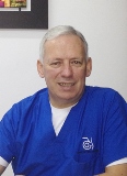
José Miguel Gómez, Profesor, MD, LO. Medical Surgeon from the Pontificia Universidad Javeriana in Bogotá, Colombia. He attended to the Century College, Minnesota for Orthotic program, and completed his orthotic residency at Gillette Children’s Hospital in Saint Paul, Minnesota. Licensed in Orthotic. He has lectured internationally.Founder and currently the Scientific Director of Laboratorio Gilete in Bogotá, Colombia and the President of Gomez Orthotic Systems, LLC Saint Petersburg FL, Assistant Professor, teaching the spinal course at Saint Petersburg College FL, North Western University IL ,South Western University TX, Pittsburg University PA, and Century College MN.Received from American Academy of Orthotics and Prosthetic, the Clinical Creativity Award 2012, in Atlanta
Abstract:
Scoliosis is a spinal curve that has more than ten degrees on the coronal plane, with a rotational component. This means scoliosis is a three dimensional deformity. To start treating a patient, the first thing needed to acknowledge is that the treatment will be done on a person, who is concerned, and is putting their health on the hands of clinicians. The treatment has to incorporate the whole patient, not the spine only. The Gomez Orthotic Spine System is a clinical protocol which measures the patient and treats them with a conservative method used for spinal deformities. The measurements, photos, and x-rays evaluated are used all in analyzing and designing the three dimensional mold. This article will demonstrate the efficacy of the system’s implementation with a single clinical case. It will focus heavily on alignment, balance, and increasing the patients stability by using the center line of each corporal plane
Mithun Neral
Case Medical Center, USA
Title: Silicone arthroplasty of the metacarpo phalangeal joint
Time : 11:45-12:10

Biography:
Mithun Neral is an orthopaedic surgery resident at University Hospitals/Case Medical Center with a particular interest in upper extremity surgery. He attended medical school at the University of Pittsburgh where he studied long-term outcomes of silicone metacarpophalangeal joint arthroplasty.
Abstract:
Abstract Introduction: Silicone arthroplasty of the metacarpophalangeal joint (MCP) is a well-established treatment for rheumatoid arthritis. However, available literature on treatment of non-rheumatic arthritis is limited to case reports and retrospective reviews of small patient populations. Purpose: The purpose of this study is to evaluate the clinical effectiveness of MCP arthroplasty for non-rheumatic arthritis in the largest group of patients with the longest follow-up period to date. We predict that MCP arthroplasty for non-rheumatic arthritis shows significant improvement in hand function, pain relief and patient satisfaction. Materials & Methods: A search of all MCP arthroplasties performed by a single surgeon for non-rheumatic arthritis over a 12-year period found 136 arthroplasties. Of these, adequate prospective follow-up assessment could be completed for 30 patients with 38 MCP arthroplasties at an average of 56 months after surgery. Objective measures included are range of motion (ROM), grip and pinch strength, disabilities of the arm, shoulder, and hand (DASH) score, and visual-analog pain score. Follow-up x-rays were reviewed. Patients also completed a subjective patient-satisfaction questionnaire. Mean ROM, DASH, and pain were compared between the pre-operative and follow-up groups by paired T-test and linear regression to identify significant differences and trends in long-term follow-up. Results: There was significant improvement between mean pre-operative and follow-up ROM, DASH, and pain with p-values of 0.0006, 0.0007 and <0.0001, respectively. Mean follow-up ROM, DASH, and pain scores were 69.5±3.0, 15.0±2.3 and 0.76±0.2, respectively. Linear regression showed significant correlations between pre-operative measurements and improvement at follow-up for ROM, DASH, and pain with p-values of 0.0003, 0.0310, and <0.0001, respectively. No significant difference existed for grip (p=0.593) or pinch (p=0.296) strength when follow-up operative and non-operative hand strengths were compared. The patient-satisfaction questionnaire showed 73% were “very satisfiedâ€, 87% would “definitely do it againâ€, and 70% experience “rare or no pain.†Follow-up x-rays showed 5° mean angulation and 2 mm mean subsidence compared to immediate post-operative X-rays. Four arthroplasties required revision, for an 11% revision rate. Conclusions: This study showed improved ROM and DASH score, excellent pain relief, and excellent patient satisfaction in patients undergoing MCP arthroplasty for non-rheumatic arthritis. Patients with more severe ROM limitation, DASH score and pain score experienced a greater improvement of these measures at follow-up. Strength improvement is limited although remains comparable to the non-operative hand. Angulation, subsidence and complications in the study population are consistent with those reported in current literature.
Ziad Elchami
International Medical Center, Saudi Arabia
Title: The effectiveness of combined radiofrequency (rf) plus epidural therapy versus epidural block alone for spinal stenosis pain due to added hypertrophied lumbosacral facets syndrome

Biography:
Ziad Elchami, Director/Consultant of the Pain and Headache Management Center at the International Medical Center (IMC), Jeddah, KSA since 2005, specializes in chronic pain and neurology; his clinical interests include clinical neurophysiology, neuromuscular disorders, and pain. Dr. Elchami completed his medical degree at the University of Damascus. He then joined Kansas University Neurology residency Program where he completed both his residency and fellowship programs in Clinical Neurophysiology Disorders. During that period, he served as Chief Resident and Chief Fellow. Dr. Elchami has a Pain and Headache Fellowship from the Cleveland Clinic Foundation where he worked from 2003 to 2005.
Abstract:
In lumbar spinal stenosis, spinal nerve roots in the lower back are compressed, producing tingling, weakness or numbness that radiates from the low back, buttocks and legs, while lumbosacral facet syndrome, a type of degenerative arthritis, occurs between the lower back and pelvis, causing significant pain throughout the lower body. The aim of the study is to evaluate the effectiveness of combined radiofrequency (RF) plus epidural therapy versus epidural therapy alone for spinal pain due to added hyperthophied lumbosacral facets syndrome. Study involved 60 patients of Pain & Headache Center, IMC, KSA. First group (N=32) underwent combined LS (RF) + epidural, applied to the lumbosacral facets region, with the following settings: 80 degrees x 1 min and repeated x 3 with repositioning of the needle. Second group (N=28) underwent epidural block only + dexamethasone. Patients were followed up to one year period. Inclusive criteria: 38 females, 22 males; ages between 40-70 years old, with mean of 38 years; and patients who already failed 9-12 sessions of ECSW or PT. Exclusive criteria: patients older than age 80; with uncontrolled diabetes and blood pressure; taking anti-coagulant; other neurological deficits; pregnant women. Average improvement of 75% for the first group, according to the numeric pain scale, was seen in patients who were treated by combined therapy; 60% in second group. Patients with low back pain who went through the combination therapy had more significant improvement than those who went through early epidural block only, with benefits lasting for more than 6 months.
P Karpe
North Tees and Hartlepool NHS Trust,UK
Title: Supramalleolar Osteotomy: A joint-preserving option for advanced ankle osteoarthritis

Biography:
Prasad Karpe has completed his Medical degree from Goa University, India. He then did his Masters of Surgery in Orthopedics from the same university. He has done 4 years of spine fellowships in India and UK. At present, he is doing a Foot and Ankle fellowship as a part of his training. He has also a member of the Royal college of Edinburgh. He has also cleared his Fellowship examinations (FRCS in Trauma and Orthopedics) and is a Fellow of the Royal college of Surgeons of Edinburgh.
Abstract:
Background: Until recently, surgical treatments for advanced ankle osteoarthritis have been limited to arthrodesis or ankle replacement. Supramalleolar osteotomy provides a joint-preserving option for patients with eccentric osteoarthritis of the ankle, particularly those with varus or valgus malaligment. Aim: To evaluate radiological and functional outcomes of patients undergoing shortening supramalleolar osteotomy for eccentric (varus or valgus) osteoarthritis of the ankle. Method: Prospective review of patients from 2008 onwards. Osteotomy was the primary surgical procedure in all patients after failure of non-operative measures. Pre-operative standing antero-posterior and Saltzman view radiographs were taken to evaluate degree of malalignment requiring correction. Radiological and clinical outcomes were assessed at 3, 6 and 12 months post-operatively. Radiographs were reviewed for time to union. Patients were assessed on an outpatient basis for ankle range of motion as well as outcomes using AOFAS scores. Results: 33 patients over a 7 year period. Mean follow-up was 25 months (range 22-30). Mean time to radiological union was 8.6 weeks (range 8-10); there were no cases of non-union. There was a statistically significant improvement in functional scoring (P<0.001); mean AOFAS score improved from 34.8 (range 15-40) pre-operatively to 79.9 (range 74-90) at 12 months post-operatively. There was no significant change in pre- and post-operative range of motion. 2 patients required revision surgery at 12 months; one to arthrodesis and one to ankle replacement. Conclusion: Supramalleolar osteotomy is a viable joint preserving option for patients with eccentric osteoarthritis of the ankle. It preserves motion, redistributes forces away from the affected compartment and corrects malalignment, providing significant symptomatic and functional improvement.
Amir A Beltagi
Cairo University, Egypt
Title: Effect of core stability exercises on trunk muscles time to peak torque in healthy adults
Time : 15:05-15:30

Biography:
Amir A. Beltagi, PT, MSc, is an Assistant Lecturer of Biomechanics, Faculty of Physical Therapy, Cairo University. He is also PhD exchange student at the Biomechanics Research Lab, Department of Orthopedics, College of Medicine, the University of Illinois in Chicago (UIC). His research project is entitled “Biomechanical changes of the spine after induced fixation: a Finite Element". Amir is an active researcher who presented his research work at many international conferences in USA, Germany, Egypt as well as published his research findings in peer-reviewed Journals.
Abstract:
Background: Core stability training has recently attracted attention for optimizing performance and improving muscle recruitment and neuromuscular adaptation for healthy and unhealthy individuals. The purpose of this study was to investigate the effect of beginner’s core stability exercises on the trunk flexors’ and extensors’ time to peak torque. Methods: Thirty five healthy individuals randomly assigned into two groups; experimental (group I) and control (group II). Group I involved 20 participants (10 male & 10 female) with mean ±SD age, weight, and height of 20.7±2.4 years, 66.5±12.1 kg and 166.7±7.8 cm respectively. Group II involved 15 participants (6 male & 9 female) with mean ±SD age, weight, and height of 20.3±0.61 years, 68.57±12.2 kg and 164.28 ±7.59 cm respectively. Data were collected using the Biodex Isokinetic system. The participants were tested twice; before and after a 6-week period during which the experimental group performed a core stability training program. Findings: Statistical analysis using the 2x2 Mixed Design MANOVA revealed that there were no significant differences in the trunk flexors’ and extensors’ time to peak torque between the “pre†and “post†tests for control group (p> 0.05). Also, there were no significant differences in the trunk flexors’ and extensors’ time to peak torque between both groups at the “pre’ test (p>0.05). Meanwhile, the 2x2 Mixed Design MANOVA revealed that there were significant differences in the trunk flexors’ and extensors’ time to peak torque between the “pre†and “post†tests for group I (p<0.0001). Moreover, there were significant differences between both groups for the tested muscles’ time to peak torques at the “post†test (p<0.0001). Interpretation: The improvement in muscle response indicated by the decrease in the trunk flexors’ and extensors’ time to peak torques in the experimental group recommends including core stability training in the exercise programs that aim to improve neuromuscular adaptation and fitness. Keywords: Core stability, Isokinetic, Trunk muscles.
Kun Wang
Southeast University, China
Title: Serum levels of the inflammatory cytokines in patients with lumbar radicular pain due to disc herniation

Biography:
Kun Wang has done his PhD in the Department of Mechanical Engineering at the Tsinghua University, China. He is now an associate professor in the Department of Radiation Oncology at Southeast University ,China.
Abstract:
Purpose: The factors influencing the presence or absence of pain in sciatica secondary to disc herniation remain incompletely understood. We hypothesized that the imbalance in inflammatory cytokines is implicated in the generation of pain. In our study, serum levels of pro-inflammatory and anti-inflammatory cytokines were investigated among patients with severe sciatica; the serum levels were compared with those of patients with mild sciatica and healthy subjects. Methods: In this prospective study, blood protein levels of the pro-inflammatory cytokines, namely, interleukin-6(IL-6), interleukin-8 (IL-8),and tumor necrosis factor-α(TNF-α), and the anti-inflammatory cytokines, namely, interleukin-4 (IL-4) and interleukin-10 (IL-10), of 58 patients with severe sciatica, 50 patients with mild sciatica, and 30 healthy control subjects were analyzed through ELISA. Physical and mental health symptoms were determined using the Oswestry Disability Index (ODI) and Short Form-36 (SF-36) questionnaire. Spearman rank correlation coefficient was also determined to calculate the correlation between the scores obtained from the questionnaires and the serum levels of cytokines. Results: No signiï¬cant difference in IL-6 levels was observed among the three groups; no signiï¬cant difference in IL-8 levels was also found between severe sciatica and mild sciatica groups, although the IL-8 levels of the healthy controls were significantly different from those of the sciatica groups(p<0.001). The TNF-α protein values were approximately fivefold higher in the severe sciatica group than in the mild sciatica group (p<0.01) and the healthy control subjects (p<0.01).The IL-4 protein levels were higher in patients with mild sciatica than in patients with severe sciatica (p= 0.05). The IL-4protein levels were also higher in mild sciatica (p=0.003) and severe sciatica (p= 0.004)groups than in the control subjects. The IL-10 protein values were higher in the severe sciatica group than in the mild sciatica group and the healthy control subjects (p<0.001).ODI was significantly correlated with IL-6 (r= 0.394, p=0.013), TNF-α (r=0.629, p<0.001), and IL-10(r= −0.415, p=0.009). By contrast, ODI was not correlated with IL-4(r= −0.174, p=0.29) and IL-8(r= −0.133, p=0.418). Conclusions: These ï¬ndings support our hypothesis that sciatica pain is accompanied by the imbalance in inflammatory cytokines.
Paul S. Sung
The University of Scranton, USA
Title: A kinematic analysis for shoulder and pelvis coordination in subjects with and without recurrent Low back pain

Biography:
Paul Sung is Associate Professor in Department of Physical Therapy at the University of Scranton, Scranton Pennsylvania. Dr. Sung conducted his research fellowship at the Iowa Spine Research Center, Biomedical Engineering Department at the University of Iowa in Iowa City, Iowa. He is a member of the International Society for the Study of the Lumbar Spine as well as the American Physical Therapy Association. His research interests include the mechanisms of chronic low back pain, sports injury mechanism, spine biomechanics, and non-operative spine care and its clinical application to neuromuscular control.
Abstract:
This presentation is to compare the shoulder and pelvis kinematics based on range of motion (ROM), angular velocity, and relative phase (RP) values during trunk axial rotation. Nineteen subjects with recurrent low back pain (LBP) and 19 age-matched control subjects who are all right limb dominant participated in this study. All participants were asked to perform axial trunk rotation activities at a self-selected speed to the end of maximum range in standing position. The outcome measures included ROM, angular velocity, and RP on the shoulder and pelvis in the transverse plane and were analyzed based on the demographic characteristics between groups. The LBP group demonstrated decreased ROM (p=0.02) and angular velocity (p=0.02) for the pelvis; however, there was no group difference for the shoulder girdle. The ROM difference between the shoulder and pelvic transverse planes had a significant interaction with age (F=14.75, p=0.001). The LBP group demonstrated a higher negative correlation between the shoulder (r=-0.74, p=0.001) and pelvis (r=-0.72, p=0.001) as age increased while no significant correlations were found in the control group. The results of this study indicated that there was a difference in pelvic rotation in the transverse plane between groups during axial trunk rotation. This pattern of trunk movement decreased due to possible pelvic stiffness with neuromuscular constraints. Since subjects with recurrent LBP demonstrated decreased pelvic rotation compared to the shoulder for postural control, increased pelvic flexibility could enhance coordinated movement patterns in order to integrate spinal motion in subjects with recurrent LBP.
Ching-Jen Wang
Kaohsiung Chang Gung Memorial Hospital, Taiwan
Title: Extracorporeal shockwave therapy for Osteoporotic Osteoarthritis of the Knee in Rats. An experiment in animals

Biography:
Ching-Jen Wang, M.D. graduated from National Taiwan University, College of Medicine. He is a board certified orthopedic surgeon and currently holds a clinical faculty at Chang Gung University College of Medicine and serves as a consultant orthopedic surgeon of Kaohsiung Chang Gung Memorial hospital, Taiwan. He has published more than 213 papers in reputed journals and has been serving as the reviewer in many journals. His primary interest and area of expertise include sports medicine, knee and hip replacement surgery, shockwave medicine and tissue regeneration.
Abstract:
The purpose of this study was to investigate the effectiveness of ESWT in osteoporotic osteoarthritis of the knee in rats. Sixty-four female S-D rats were divided into four groups. The control group received sham ovariectomy (OVX) and sham anterior cruciate transection (ACLT) and medial meniscectomy (MM). The osteoarthritis (OA) group received ACLT and MM, but no OVX. The osteoporosis (OP) group underwent bilateral OVX, but no ACLT and MM. The OA + OP group received bilateral OVX, ACLT and MM. One half of animals also received ESWT. The evaluations included the areas of gross pathology, bone mineral density (BMD), micro-CT scan, bone strength test, histopathological examination and immunohistochemical analysis. Group OA + OP showed larger areas of arthritic changes than groups OA and OP as compared to sham group. BMD and bone strength significantly decreased in groups OA, OP and OA+OP versus sham group, and ESWT significantly improved the changes. In micro-CT scan, subchondral plate thickness significantly decreased and bone porosity increased in groups OA, OP and OA+OP, and ESWT significantly improved the changes. .Mankin and Safranin O scores significantly increased in groups OA and OA+OP, relative to sham group, and ESWT significantly improved the changes,. DKK-1 significantly increased, and VEGF, PCNA and BMP-2 decreased in groups OA, OP and OA+OP relative to sham group, and ESWT significantly reversed the changes. This study showed that osteoporosis increased the severity of osteoarthritis of the knee. ESWT was shown effective to ameliorate osteoporotic osteoarthritis of the knee.
Rachel W. Li
Australian National University Medical School, Australia
Title: Heparanase may have a key role in the regulation of inflammatory mediators in Rheumatoid Arthritis

Biography:
Rachel W. Li (MD, PhD) completed her PhD in Australia and gained her postdoctoral experiences in molecular pharmacology focusing on immune regulation of metabolic diseases at the University of Hawaii, the USA. She returned to Australia joining the Trauma and Orthopaedic Research Unit (TORU) at the Australian National University Medical School and has established TORU Laboratory. She is currently leading her team with a focus on osteoimmunology and biomaterials in bone remodeling.
Abstract:
Heparanase is the only known mammalian endoglycosidase capable of degrading the heparan sulfate (HS) glycosaminoglycan, both in extracellular space and within the cell. HS is reported to control inflammatory responses at multiple levels, including the sequestration of cytokines/chemokines in the extracellular space, the modulation of the leukocyte interaction with the endothelium and ECM, and the initiation of innate immune responses. We have reported heparanase expression in synovium of rheumatoid arthritis (RA) patients, and this new finding may offer a new insight of the potential regulatory role of heparanase in the disease activity of RA.However, the precise mode of action by heparanase in inflammatory reactions of RA remains largely unknown. The aim of this project was to examine the heparanase activity, its expression and correlation with the inflammatory mediatory and angiogenic gene expression in plasma and synovium of RA patients, with an ultimate goal of developing heparanase as a potential predictor of RA progression and a new therapeutic target. We have found that a highly significant increase of heparanase activity and expression in synovial fluid and synovial tissue of RA patients, and an increase of the heparanase activity positively correlate with the inflammatory and angiogenic gene expression. We also have some evidence to support a postulation that the involvement of heparanase in gene regulation in the development of pannus in RA may be reflected in a patient’s blood, thus heparanase can be a potential predictor of RA progression and a novel therapeutic target.
Yves Lignereux
Natural History Museum & National Veterinary College, France
Title: Vertebral ankylosing hyperostoses in the horse in archeological contexts: differential diagnosis and comparative nosological review

Biography:
Yves LIGNEREUX, DVM, PhD, Agrege, is Professor of anatomy at the National Veterinary College, Toulouse, France. He has specialized in archeozoology and has been in charge of the scientific programme for the refoundation of the Natural History Museum of Toulouse, reopened to the public in 2008. His current projects in archeozoology are in France, Spain, Cyprus, Egypt, Mongolia.
Abstract:
In the veterinary clinical literature, two basic forms of vertebral fusions have been described: spondyl(arthr)itis/-osisankylopoetica (SPA) and spondylitis/-osischronicadeformans (SPD)(SILBERSIEPE and BERGE, 1958; MORGAN, 1967);advances in clinics have added DISH as a third form, principally in dogs (WOODARDet al., 1985). Inarcheo(zoo)logical contexts, SPA refers to bony bridges connecting synovial joints, while SPD refers to bony bridges between adjacent vertebral bodies(BARTOSIEWICZ, 2013). In human clinics and paleopathology, SPA, SPD and DISH are well-defined entities (e.g. ADLER, 2000; ORTNER, 2003; PAJA, 2012), but with lesional expressions specifically different from animals’. SPA and DISH both are inflammatory, productive, enthesopathic/periosteal connective tissue conditions; SPD is degenerative, erosive, and osteo-/syndesmophytic. SPA is characterized by dorsal and lateral fusions and somatic false-ankyloses; SPD is signaled by spinouspseudarthroses, small-joint osteoarthritis, and disk disease. In all spondyloarthropathies, the ventral longitudinal ligament is highly reactive. Ancient domesticated horses often show lesions of vertebral ankylosing hyperostosis diagnosed as SPA when affecting vertebral bodies and SPD when affecting dorsal arches and their processes. But both processes can interact in a mechanical action-reaction spiral, so that they often coexist on the same vertebra(LEVINEet al., 2005), even questioning the very meaning of differentiating two entities(THILLAUD, pers. com.).Thus, the main supposed etiology is mechanical stress and the causes are looked for in the way horses were handled and utilized. Discussing published cases gives the opportunity to a comparative and critical review of spinal hyperostosing and ankylosing diseases in horse and man.

Biography:
Ole Kudsk Jensen, a specialist of Rheumatology and since 2004 leader of a multidisciplinary research unit of the Spine Center of Silkeborg Regional Hospital, Central Region Denmark. In 2009, he completed his PhD from the University of Aarhus about sensitization of the nociceptive system in low back pain patients. He has published more than 25 papers in international journals, and 12 of these articles have been about sick-listed low back patients. He has been a member of the national Danish board working out guidelines for referral for back surgery.
Abstract:
In 325 sick-listed low back pain (LBP) patients, a multivariate prognostic model for unsuccessful return to work (U-RTW) was developed and validated with satisfying result in a subsequent cohort of 120 patients. These patients were recruited, randomised and managed in the same way as the former group. U-RTW was not different in patients with and without radiculopathy. The model included back+leg pain intensity, side-flexion, bodily distress and four psychosocial risk factors. Magnetic resonance imaging of the lumbar spine was performed consecutively in 141 of these patients. The degenerative manifestations were described standardised, including disc herniations and nerve root compromise. Only type1 Modic changes identified in 18% were negatively associated with U-RTW, also after adjustment for already established risk factors.
Oslei de Matos
Federal Technological University of Parana, Brazil
Title: Influence of sleep disorders on body composition in women with Fibromyalgia
Biography:
Oslei de Matos has completed his PhD in Education and Sports Science at the age of 41 years from Porto University and master degree in Physical Education and Physiotherapy studies from Pontifical Catholic University of Paraná in 1989 and 1995. Medical Gymnastic Specialist in Castelo Branco University of Rio de Janeiro in 1991. He is the director of Laboratory Biochemical and Densitometry, Researcher on women’s health with Osteoporosis, Fibromyalgia and Manager. He is Author of books on postural and physical education. Currently is Anatomy and Biomedical Professor in Graduate Program in Biomedical Engineering in Federal Technological University of Parana in Curitiba – Brazil.
Abstract:
Background: The main objective of this research is relation between sleep disturbance and body composition in women with fibromyalgia. This study used DXA to body composition and Pittsburgh sleep quality index to analyze the sleep quality. This research showed that sample has presence of sleep disturbance with low quality of life, moderate pain. These data showed the relation with excess of fat mass and calories intake. The polysomnography it use to analyze the quality of sleep and to DXA body composition. These data define at what stage of sleep is most affected and the physiological consequences entailed by such phases. Through this analysis correlates with memory loss and alteration of muscle and a greater tendency to fatness components. The main causes of sleep apnea and hypopnea refer to central adiposity, and this will be reviewed after bariatric surgery and improving the quality of sleep after. Other resources used for the diagnosis of fibromyalgia are the Thermography and digital photography. The main objective is the correlation of the pain points with the thermographic analysis of the regions of greatest cellular activity of women with fibromyalgia. The standard are used for the analysis of generalized pain, pain intensity, and quality of life. As the obesity has relation with fibromyalgia, the nutritional assessment it is essential for control of confounding variables in the disease, as well as adaptation to diet for likely improvement of metabolism with increase of antioxidant capacity and facilitation of sleep regulation. All these data are shared with specialized team medical to pharmacological adaptation.
Moin Khan
McMaster University, Canada
Title: Arthroscopic Surgery for Degenerative Tears of the Meniscus: A Meta-Analysis
Biography:
Dr. Khan completed his undergraduate education in the Bachelor of Health Sciences program at McMaster University graduating Summa Cum Laude. He went on to complete his medical training at Michael G. DeGroote School of Medicine in 2010 and graduated from McMaster University's Orthopaedic Surgery Residency Program in 2015. As a recipient of the Margaret and Charles Juravinski Surgical Fellowship award he concurrently completed a Master’s Degree in Health Research Methodology with a specific research focus on clinical outcomes research under the supervision of Dr. Mohit Bhandari and Dr. Olufemi Ayeni. Dr. Khan's research and subspecialty interests relate to orthopaedic sports medicine and in particular the rapidly expanding field of hip preservation surgery. He has published numerous peer-reviewed papers, received national and international media attention for his research, presented at national and international conferences and received multiple research awards for his work.
Abstract:
Introduction: Partial meniscectomy is one of the most commonly performed orthopaedic procedures. Controversy exists with regards to its efficacy in the setting of degenerative meniscal tears. This systematic review and meta-analysis evaluates the efficacy of arthroscopic partial meniscectomy in the setting of mild to no concurrent osteoarthritis of the knee in comparison to non-operative or sham treatments. Methods: Two reviewers independently screened MEDLINE, EMBASE, PUBMED and Cochrane databases for all eligible studies. Only randomized control trials published from 1946 through to Jan 2014 were included. A risk of bias assessment was conducted for all included studies and outcomes were pooled using a random effects model. Outcomes were dichotomized to short-term (< 6 months) and long-term (< 2 years) data. The quality of the evidence was assessed and confidence in recommendations was developed according to the Grading of Recommendations Assessment, Development, and Evaluation (GRADE) approach. Results: Seven randomized control trials were included in this review. Six of the seven included studies had moderate to significant risk of bias. The pooled treatment effect across studies did not demonstrate a statistically significant or minimally patient important difference (MID) between treatment arms for functional outcomes (SMD 0.07, 95% CI -0.10 to 0.23, p=0.43) (I2=20%) or pain relief (MD= -0.06, 95% CI -0.28, 0.15, p=0.57, I2=0%) at two years. Short-term functional outcomes between groups were statistically significant but did not exceed the threshold for MID. (SMD 0.25, 95% CI 0.02 to 0.48, p=0.04). (I2=56%) A priori subgroup analysis related to conservative treatment resulted in substantial decrease in heterogeneity. Sensitivity analyses related to sample size and missing data did not have a significant effect on treatment effect. Conclusions: According to the GRADE approach, there is moderate evidence to suggest there is no benefit of arthroscopic partial meniscectomy for degenerative meniscal tears in comparison to nonoperative or sham treatments for individuals with mild or no concomitant osteoarthritis. A trial of non-operative management should be the first line treatment in such individuals. Future research into identifying indications and ideal patient selection is required with regards to prognostic factors, which may influence outcomes following surgical and conservative treatment.
Abdulkarim Ali
Midland Regional Hospital, UK
Title: Agricultural and Farming Injuries in the Irish Midland
Biography:
Abdulkarim Ali is working in Midland regional hospital, Tullomore, Ireland, UK. He has a interest in Bacterial contamination of Diathermy tips, Intrinsic properties of surgical gloves and Total knee Arthroplasty. He completed his studies from University of Limerick, Ireland, UK
Abstract:
Background: The agricultural and farming business are an important source of employment in the Midlands. This is a retrospective study examining the demographics, characteristics, and outcomes of agricultural and equestrian related injuries presenting to the Midland Regional Hospital, Tullamore, Co. Offaly. Methods: Every presentation to the Accident & Emergency Department at the Midlands Regional Hospital in 2013 was assed retrospectively to determine if an injury had been sustained in an agricultural or equestrian environment. Patient characteristics and injury details were collected for 345 patients who attended the Accident & Emergency Department. Patient demographics, month of occurrence, mechanism of injury, radiology results, management and follow up data were collected and analysed. Results: There were 196 agricultural related presentations to the Accident & Emergency Department and 149 equestrian related presentations. 23% of the agricultural injuries and 36% of the equestrian injuries had confirmed radiological evidence of a fracture or joint dislocation. There were significantly more males involved in agricultural injuries than females (98% vs 2%, p < 0.001). There were significantly more females involved in equestrian injuries than males (58% vs 42%, p < 0.05). 10% of farming injuries and 15.4% of equestrian injuries required admission. Farming machinery accidents contributed to significantly more admissions than any other cause in the agricultural category (p < 0.01). Conclusion: agricultural and farming related injuries in the Irish midland are common presentations to orthopaedic surgeon. Increased attention to occupational health hazards seems required in the equestrian environment as Prevention of adverse health outcomes.
- Ultrasound, Imaging and Diagnostic Techniques
Orthopedic Surgery and Orthopedic Trauma
Musculoskeletal Outcomes: Theory and Practice
Session Introduction
Lloyd S Miller
Johns Hopkins University, USA
Title: In vivo bioluminescence, fluorescence, µCT and photoacoustic imaging tononinvasively evaluate pathogenesis, therapeutics and diagnosticsin an experimentalorthopaedic implant infection
Time : 10:10-10:35

Biography:
Miller completed his M.D. and Ph.D. degrees from SUNY-Downstate Medical Center and his clinical residency and post-doctoral fellowship training at UCLA School of Medicine. He currently is an Associate Professor in the Johns Hopkins Departments of Dermatology, Medicine (Division of Infectious Diseases) and Orthopaedic Surgery. He has published over 30 peer-reviewed manuscripts, including high impact publications in Immunity and Journal of Clinical Investigation. For more than 10 years, Dr. Miller has been a pioneer in utilizing in vivo whole animal optical (bioluminescence and fluorescence imaging), µCT and photoacoustic imaging to study infectious diseases, including orthopaedic implant infections.
Abstract:
Infection isa devastating complication following orthopaedic surgical procedures.Bacteria form biofilmson the implanted materials,creatingchronic infection and inflammation that leads to periprosthetic osteolysis, implant loosening and failure.These infections are challenging to diagnose and treatment is difficult and typically requiresreoperations, prolonged systemic antibiotic courses, extended disability and rehabilitation. To study the these infections, we developed a mouse model of an orthopaedic implant infectionin which a bioluminescent strain of Staphylococcus aureus was inoculated into a post-surgical knee joint of LysEGFP mice (expressing fluorescent neutrophils) that contained a titanium Kirschner-wire extending from the femur. In vivo bioluminescence and fluorescence imaging was used to noninvasivelyquantify the bacterial burden andneutrophil recruitment, respectively. We found that bioluminescent bacteria persisted for the 48 day experiment whereas EGFP-neutrophil signals decreasedwithin the first 14-21 days. Using µCT imaging, the outer bone volume of the femur doubled, which was associated with low-density signals and bone loss, consistent with peri-implant osteolysis.This model was used to evaluate optimal antibiotic combinations to treat an existing infection as well as the novel antimicrobial implant coatings. Finally,as a new diagnostic approach, near-infrared fluorescent probes were used in combination with advanced techniques of photoacoustic imaging. This imaging modalityhas the potential for clinical translation since photoacoustic signals can be detected several centimetersinto tissue. Taken together, bioluminescence, fluorescence, µCT and photoacoustic imaging represent valuable noninvasive techniques to study the pathogenesis of orthopaedic implant infections and to evaluate novel therapeutic and diagnostic strategies.
Artur Yudi Utino
Federal University of Sao Paulo, Brazil
Title: Correlation of the degree of clavicle shortening after non-surgical treatment of midshaft fractures with upper limb function
Time : 10:35-11:00
Biography:
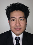
Artur Yudi Utino has completed his medical degree at Universidade Federal de São Paulo, Brazil, in 2011. Concluded his residency in orthopaedics surgery in 2015 and currently is doing a fellowship on Shoulder and Elbow Surgery. Completed his master’s degree in 2015 at the same university
Abstract:
Background: Despite the use of non-surgical methods to treat for the majority of mid-shaft fractures of the clavicle, it is remains controversial whether shortening of this bone following non-surgical treatment of a middle third fracture affects upper limb function. Methods: We conducted a cohort study by sequentially recruiting 59 patients with a fracture of the middle third of the clavicle. All patients were treated non-surgically with a figure-of-eight bandage until clinical and radiological findings indicated healing of the fracture. Functional outcome was assessed using the Disability of Arm, Hand and Shoulder (DASH) score revalidated for the Portuguese language, other outcomes assessed included: pain measured by visual analogue scale (VAS); radiographies to measure the degree of shortening, fracture consolidation and fracture mal-union. Information was also collected regarding the mechanism of injury, patient’s daily activities level and epidemiological features of the patient cohort. The results of our findings are expressed as the comparison of the functional outcome with the degree of shortening. Results: Patients were assessed six weeks and one year after injury. In the first evaluation, the mean DASH score was 28.84 and pain measured by VAS was 2.57. In the second evaluation (one year after injury) the mean DASH score was 8.18 and pain was 0.84. The mean clavicle shortening was 0.92 cm, ranging from 0 to 3 cm (SD=0.64). There was no correlation between the degree of shortening and DASH score after six weeks and one year (p=0.073 and 0.706, respectively). When only patients with of shortening greater than 2 cm were assessed for correlation, the result did not change. Conclusion: We conclude that clavicle shortening after nonsurgical treatment with a figure-of-eight bandage does not affect limb function, even when shortening exceeds 2 cm.
Navtej Sathi
Forth Valley Royal Hospital, UK
Title: Musculoskeletal aspects of hypoadrenalism: Just a load of aches and pain?
Time : 11:20-11:45

Biography:
Sathi is currently working as a consultant in Acute Medicine in Scotland. Previously he has previously held posts as a Consultant in Acute Medicine and Rheumatology. His interests include pain, integrated medicine, rheumatology, methotrexate side effects and interstitial lung disease. He holds an M.Sc. in Pain Management from Cardiff University. His research governance score (from Research Gate) is above 20, placing him in the top 30 percent in the world. His talk on musculoskeletal aspects of hypoadrenalism may provide some insight into how chronic disease can give rise to musculoskeletal symptoms and signs
Abstract:
Hypoadrenalism remains an underdiagnosed disease process. It’s musculoskeletal features are less well described in the literature when compared to diabetes or thyroid disorders. As a consequence considering hypoadrenalism in a patient with mainly musculoskeletal features may be difficult and delayed. The paper will include three cases from our own experience as well as all the relevant cases of hypoadrenalism in the English, French and Spanish language medical literature, going back to the time of Thomas Addison. There will be a summary of the relevant signs, symptoms and investigation findings which may help clinicians diagnose and treat this disease. Also the relationship of hypoadrenalism and chronic disease may be explored during the talk.
Renato A Sernik
Sirio Libanes Hospital, Brazil
Title: Update on the use of ultrasound in the diagnostic of carpal tunnel syndrome
Time : 11:45-12:10

Biography:
Renato A Sernik is a Doctor and obtained his PhD from the University of Sao Paulo. He is a Radiologist at the Medical School Hospital of the University of SaoPaulo for 16 years. He is the author of some books “Ultrasound of the Musculoskeletal System (1999) and Ultrasound of the Musculoskeletal System-Correlation with MRI (2009)”, translated into Spanish and Italian. He is working as a Radiologist at the Sirio Libanes Hospital from 14 years.
Abstract:
Ultrasonography has become one of the main complementary tests for the diagnosis of carpal tunnel syndrome, especially in patients with a history of diseases that may be associated with peripheral neuropathy, such as diabetes mellitus, or in cases where the electroneuromyography is doubtful. The method has also been useful in patients with unsatisfactory results after surgical treatment, where fibrosis, adhesions and neuromas can be identified, having the advantage compared to MRI to be dynamic examination and lower cost. However, the method is operator-dependent with a longer learning curve. Between 2006 and 2014 we analyzed about 120 wrists of patients with clinical suspicion of carpal tunnel syndrome, with electroneuromyographic correlation, being part of the results published in the journal Skeletal Radiology (2008). The increase in cross-sectional area associated with the change in echotexture of the median nerve are the main criteria used for the diagnosis of carpal tunnel syndrome.
Adham Elgeidi
Mansoura University, Egypt
Title: Interleukin-6 and other inflammatory markers in diagnosis of periprosthetic joint infection
Time : 12:10-12:35

Biography:
Adham Elgeidi has completed his MD at the age of 35 years from Mansoura University and postdoctoral studies from both Mansoura University School of Medicine, Egypt and Peninsula University School of Medicine, UK. He is currently a consultant and assistant professor of Orthopaedics and Trauma with special interest in sports medicine and arthroplasty. He is the director of Orthopaedics and Trauma undergraduate teaching and training program Mansoura University. He has published more than 20 papers in reputed journals and has been serving as an editorial board member of repute. He has supervised more than 15 MSc & MD thesis. He has presented more than 30 papers in different conferences. He used to work in Plymouth, UK (May 2003 to August 2006), Tuebingen, Germany (September 2005 to October 2005) and Bordeaux, France (March 2007).
Abstract:
Purpose: to evaluate diagnostic value of interleukin- 6 (IL-6) and other inflammatory markers; C- reactive protein (CRP), Erythrocyte sedimentation rate (ESR), and White blood cell count (WCC) in diagnosis of PJI. Methods: Study group included 40 patients (21 males, 19 females) admitted for surgical intervention after knee or hip arthroplasties. Patients were subjected to careful history taking, thorough clinical examination and preoperative laboratory investigations including serum IL-6, CRP, WCC and ESR. Peri-implant tissue specimens were subjected to microbiological culture and histopathological examination. Results: Mean age of studied patients was 58.4 years (range, 38-72 years). Intraoperative cultures and histopathological examination revealed 11 patients had been infected (PJI) and 29 patients were aseptic failure of prosthesis. Four presumed markers of infection were tested preoperatively: ESR; CRP; WCC; and IL-6. ESR (p=0.0001), CRP (p=0.004), WCC (0.0001), and IL-6 (p=0.0001) were significantly higher in patients with septic revision than those with aseptic failure of the prosthesis. Serum IL-6 (> 10.4pg/ml) reportedly had a sensitivity (100%), a specificity (90.9%), a PPV (79%), a NPV (100%), and accuracy (92.5%). Conclusions: The present study demonstrated that IL-6 has been found to be the most accurate laboratory marker for diagnosing PJI when compared to ESR, CRP, and WCC. IL-6 above 10.4 pg/ml and CRP level above 18 mg/L will identify all patients with PJI and the combination of CRP + IL-6 is an excellent screening test to identify all such patients (sensitivity 100%, NPV 100%).
Tesfaye Hordofa Leta
University of Bergen, Norway
Title: The outcome of unicompartmental knee arthroplasties after aseptic revision into total knee arthroplasties
Time : 12:35-13:00

Biography:
Tesfaye Leta received BEd in Pedagogical Sciencefrom Bahir Dar University, Ethiopia in 1998, BSC inNursing from Bergen University College in 2008, andMPhil in International Health from University of Bergen in 2010. He is working as authorized nurse at the department of orthopeadic surgery, Haukeland University Hospital since 2010: Currently he is a PhD candidate atthe University of Bergen, Bergen,Norway since 2013. The title of his PhD project is Outcome of revision knee arthroplasty in Norway (1994-2011) with special focus on implants survival rate, risk of re-revision, pain, satisfaction, and health related quality of life.
Abstract:
Background: The general recommendation for a failed primary unicompartmental knee arthroplasty (UKA) is revision to a total knee arthroplasty (TKA). Many surgeons prefer to use UKAs in younger patients to postpone TKA believing that the results after revision from a UKA to a TKA is equal to a primary TKA and better than a revision TKA. For this to be true the rev-UKA should outperform the rev-TKA. The purpose of the present study was to compare the outcomes, surgical procedures, and mode of failure of failed primary UKAs and primary TKAs revised to TKAs. Method: The study was based on 768 failed primary TKAs revised to TKAs (rev-TKAs) and 578 failed primary UKAs revised to TKAs (rev-UKAs) and reported to the Norwegian Arthroplasty Register between 1994 and 2011. Patient-reported outcome measures (PROMs) including the EQ-5D, the Knee Injury and Osteoarthritis Outcome Score (KOOS), and Visual Analogue Scales assessing satisfaction and pain were used. Kaplan-Meier and Cox-regression analyses were performed to assess the survival rate and the risk of re-revision. The independent student’s t-test and multiple linear regression were used to estimate the differences in mean scores in PROMs between the two groups. Results: Overall, 12% of rev-UKAs and 13% of rev-TKAs were re-revised between 1994 and 2011. The 10 years survival percentage of rev-UKAs vs rev-TKAs was 82 vs 81%, respectively (p=0.1). There was no difference in the overall risk of re-revision for rev-UKAs vs rev-TKAs (RR= 1.3; p=0.1), nor did we find any differences in the PROM scores. However, the risk of re-revision was 2 times higher for rev-TKA patients aged over 70 years (RR= 2.2; p=0.04). Loose tibia (28 vs 17%), pain alone (22 vs 12%), instability (19 vs 19%), and deep infection (16 vs 31%) were major causes of re-revision for rev-UKAs vs rev-TKAs, respectively but the observed differences were not statistically significant. The surgical procedure for rev-TKAs took longer time (mean=150 vs 114 minutes) and more of the operations needed stems (58 vs 19%), and stabilization (27 vs 9%) compared to rev-UKAs. Conclusion: Despite the surgical procedure of rev-TKA seeming technically more difficult compared to that of rev-UKA, the mode of failure and the overall outcome of rev-UKAs and rev-TKAs were similar. Thus, the argument that the outcome of revising UKA to TKA is similar to primary TKA could not be supported by the present study findings.
Cary Fletcher
St. Ann’s Bay Hospital, Jamaica
Title: Open locked nailing using an expandable nail: An alternative approach
Time : 13:50-14:15

Biography:
Cary Fletcher completed his MBBS degree at University of the West Indies in 2001 and went on to complete his Doctorate in Orthopaedics in 2012, also at The University of the West Indies at the age of 37 years. He is now an Orthopaedic surgeon at St. Ann’s Bay Regional Hospital where he functions as the academic coordinator of the Orthopaedic service. As coordinator, he functions to moderate presentations at all levels of medical staff including residents, interns and student nurses and hence ensure continuing up to date medical education. Doctor Fletcher serves as one of the senior surgeons on the service in which the majority of cases involve Orthopaedic trauma. He is first author on two publications, fifth author on a third and first author on a case report which has been accepted for publication.
Abstract:
Objective: The main objective is to evaluate various outcomes of open intramedullary nailing using the Fixion expanding nail at our institution. Method: A retrospective study was performed using the hospital records. The mechanism of injury, the time between injury and surgery, blood transfusion requirements, blood loss, surgical times, time taken to weight bear (for the femoral/tibial fractures), time for commencement of upper limb use (for humeral fractures), complication rates and the average follow up times were documented. Fifty-seven Long bone fractures in 57 patients were included in this study. Complete results including preoperative X-Rays were available for 27 patients. In 30 cases, the actual X-Rays were not located but documentation by the treating surgeons was available. Results: There were 44 acute femoral fractures, 6 acute tibial fractures, 3 acute humeral fractures, 2 humeral nonunion, 1 tibial nonunion and 1 pathological femoral fracture. All patients achieved radiological union and the complication rates were deemed acceptable. Conclusion: Open intramedullary nailing using an expanding nail may be used for a variety of indications involving the humerus, tibia and femur.
Tariq Alotaibi
King Saud University, Saudi Arabia
Title: Thoracic vertebral hemangioma causing lower limb spastic paresis (case report)
Time : 14:15-14:40
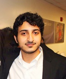
Biography:
Tariq alotaibi : medical student in the final year from king saud university ,riyadh saudi arabia . He is interested in orthopedic. Working on two different research projects in orthopedic.
Abstract:
In this paper we report a rare case of aggressive vertebral hemangioma in an 18-year old male presenting with paraparesis, disturbed sensations and spasticity. Vertebral hemangioma can present with wide variety of symptoms based on extension and compression of the spinal cord. It’s more common in adult. Radiological Imaging were diagnostic of vertebral hemangioma in the level of T8 involving the whole vertebral body. Patient was treated surgically and recovered completely. The clinical presentation, finding on radiological studies, and the management are discussed below.
Abed Al-Negery
Mansoura University, Egypt
Title: Vascularized fibular medialization for reconstruction of the tibial defects following tumor excision
Time : 14:40-15:05

Biography:
Abed Abdel Latif Mohammed Al Negery is a medical Practitioner at Mansoura University hospital and Abed Orthopedic Center, Egypt. He is a professional member of Egyptian Orthopaedic Association and Egyptian Oncology Association.
Abstract:
Purpose: The purpose of this study was to evaluate the functional and oncologic results of fibular medialization when used along as a single-stage reconstructive technique after wide excision of malignant tumors of proximal, middle or distal tibia. Methods: Between December 2010 and May 2015, 14 patients (6 males and 8 females) with primary malignant tumors of the tibia (8 proximal, 4 diaphyseal, 2 distal) were treated by wide excision. The mean age of the patients at the time of surgery was 23.2 years (11 to 38). The fibula was mobilized medially with its vascular pedicle to fill the defect. The average size of the defects reconstructed was 19.5 cm (18 to 22). Full weight bearing was achieved at 6.2 months (range: 4 to 10) postoperatively. Patients were evaluated functionally using the Musculoskeletal Tumor Society evaluation system. Results: The mean follow-up period was 31.3 months (range: 17 to 54). The average time for complete union was 6.2 months (range: 4 to 10). At final follow up all patients had fully united grafts; 9 walked without aids. Chest metastases developed in one patient, superficial wound infection in one patient and leg length discrepancy in three patients. The mean Musculoskeletal Tumor Society Score was 23/30 points (76.5%). The minimum score was 40% and the maximum was 90%. Conclusion: Ipsilateral pedicled vascularized fibular centralization or medialization is a durable reconstruction for tibial defects after wide excision of bone tumors with an acceptable functional outcome. Stable osteo-synthesis is the key to union.
Garima Gupta
Saaii College of Medical Science and Technology, India
Title: Prevalence of musculoskeletal disorders in farmers of Kanpur-rural, India
Time : 15:05-15:30

Biography:
Garima Gupta is result oriented physiotherapist. She is presently working as a Head of Department, Researcher and Assistant Professor in Saaii College of Medical Science and Technology, India. She has done her graduation from super specialty HOSMAT hospital Bangalore. In 2010 she completed her Masters in Physiotherapy (Neurology) from Indian Spinal Injury Center, New Delhi. She is actively involved in various ongoing research projects and has multiple international books and research publication in the field of physiotherapy. She actively contributes as reviewer for many international journals. She has also presented two papers in international conferences
Abstract:
Background: Musculoskeletal Disorders are prevalent and the impact is pervasive across a wide spectrum of occupations, as is evident from numerous studies conducted across the globe. However, there are very few studies that document the prevalence of MSDs in India, and there are hardly any studies that focus on the country’s farming community, which constitutes more than 58 percent of the Indian work force. Thus in the present study an attempt has been made to analyze the prevalence of MSDs in farmers of Kanpur-Rural, India. Methods: A sample of 300 farmers of Kanpur rural district, aged between 20-70 years, was selected. NMQ to measure the musculoskeletal disorders was given to all the farmers. Results: Analysis of data identified four most common MSD affecting the farmers of Kanpur-Rural: LBP (60%); knee pain (39%), shoulder pain (22%), and neck pain (10%). Conclusion: Finding of the present study shows that yearly prevalence of MSDs in farmers of Kanpur-Rural, India is alarmingly high and it suggests that nearly 60 percent of Indian cultivators could be afflicted by this disease, which urgently needs to be corroborated by similar studies at the national level. LBP is the most prevalent type of MSDs affecting the famers. Knee, shoulder and neck pain are other important MSDs affecting farmers in the study area.Observations made during the present study suggest that poor postures and lack of ergonomic awareness in the farming community are the two principal causative factors contributing to the development of MSDs.
Sameer M Naranje
Helena Regional Medical Center, USA
Title: An update on lower limb joint replacement in rheumatoid arthritis

Biography:
Sameer Naranje has completed his fellowship in joint replacement and adult reconstructive surgery from University of Minnesota after completing his M.R.C.S from Royal College of Physicians and Surgeons, Glasgow and Orthopaedic Residency from prestigious All India Institute of Medical Sciences, New Delhi, India. He is currently Chief of Surgery and Orthopaedics at Helena Regional Medical Center, AR, USA and Consultant Orthopaedic Surgeon at East Arkansas Orthopaedic Associates, AR, USA. He has extensive research background, numerous conference presentations, has published more than 30 papers in reputed journals and has been serving as an editorial board member and reviewer of number of reputed Orthopaedic journals.
Abstract:
More than 2/3rd of patients of Rheumatoid arthritis (RA) become disabled 20 years from primary diagnosis. RA is one of the most common indications for lower limb joint replacement in Northern Europe and North America. Though improved medical treatment of RA over last 2 decades have decreased the rate of hip and knee surgery, over a third of patients will need a major joint replacement, of which the majority will receive a total hip or knee replacement (THR and TKR). This paper summarizes an update on the major lower limb arthroplasty surgery for patients with RA. A multidisciplinary approach is needed for preoperative optimization. RA patients may need joint replacement at relatively younger age when compared to the patients with osteoarthritis and may need mutilple revision surgeries over their lifespan. Patients should be made aware of this and increased risk of infection and periprosthetic fracture rates associated with their disease. Biologic agents should be stopped pre-operatively due the increased infection rate. However, continued use of methotrexate does not increase infection risk, and may infact be helpful in recovery. The surgical sequence is commonly hip, knee and then ankle. Cemented total hip replacement (THR) and total knee replacement (TKR) have superior survival rates over uncemented components. Achieving ligamentous balance may be challenging in knee replacements in these patients and more constrained implants may be needed in patients with poor ligaments and severe deformities. The evidence is not clear regarding a cruciate sacrificing versus retaining in TKR, but a cruciate sacrificing component limits the risk early instability and potential revision. The results of total ankle replacement remain inferior to THR and TKR though the science of ankle replacement continues to evolve. RA patients achieve equivalent pain relief after joint replacement, but their rehabilitation is slower and their functional outcome may not be optimum due to continued presence or worsening of the disease. Again, the key to managing these complicated patients is to work as part of a multidisciplinary team to optimize their outcome
Andrew Clerk
Ayr University Hospital, UK
Title: A series of nerve repairs on traumatic nerve injuries
Biography:
I am Andrew Clerk and I work as a Speciality Trainee in orthopaedics. I have been working in orthopaedics for more than 5 years. My qualifications are MB ChB and MRCS.
Abstract:
Nerve injuries are a difficult surgical problem to tackle with. They have been accepted as a entity with poor prognosis. We share our experience of surgical repair of nerve injuries in three different hospitals of South Asia and Western Europe. The objectives of this retrospective study was to audit the results of nerve repairs done within a period of 10 years in three different centres of the world and assess the outcome. A retrospective analysis of the nerve repairs of the upper limb as well as of the lower limb of 456 patients were under taken in this study. The study period was 10 years from January 2003 to October 2013 and involved nerve repairs done in three institutions. Case notes of nerve repairs identified from theatre registers were used to gain information of the relevant patients. Pre-operative assessment notes and operative notes were used to identify the nerves involved, the mechanism of injury and operative interventions undertaken. Follow-up details were obtained also from the records in the case notes of the patients. A total of 456 patients were looked at in three different hospitals in Colombo, Manchester and Ayr. Out of the 456 patients 382 were males and nearly 68% were due to violence. About 12% were due to domestic accidents and 20% were occupational injuries. Out of the nerve injuries 76% were involving the upper limb and a total of 79% were associated with fractures. Three hundred and twenty one patients had primary nerve repairs. Out of the patients who had primary nerve repairs nearly 62% achieved a good functional out come. Of the group of patients who had delayed nerve repairs due to various reasons only 23% achieved a good functional out come. Common causes for nerve injuries are related to violence and occupational hazards. They are more common in the upper limb. Nerve injuries are best dealt with in the primary wound exploration where ever possible for best functional out come. Delayed exploration and repair is not entirely without a good functional result.
Thisara C Weerasuriya
Tameside General Hospital, UK
Title: Sub-laminar wiring in spinal fractures – A viable alternative

Biography:
I have gained experience in general orthopaedics, in trauma as well as in elective orthopaedic practice both in Sri Lanka and in the United Kingdom. My practice in orthopaedics spans nearly ten years from the year 2000 to date of which the majority of my experience was gained in my native country, Sri Lanka. Inaddition to primary and revision joint replacements I am specially interested in instrumentation of traumatic spinal fractures and limb salvage surgery. Specialties:My special interests are Limb salvage and trauma. In the field of limb salvage surgery I have expeprience in salvage and compartmentectomy of limbs involved in bone tumours with allograft recontruction. In the field of trauma my special interest is on salvaging limbs deamed beyond salvage by most salvage scoring systems with emphasis on re-implantation of severed limbs due to traumatic amputations. My current series is 36 limb re-implantations during the past 10 years.
Abstract:
Introduction In the coastal regions of the island nation of Sri Lanka (Ceylon) toddy tapping form coconut trees is a common occupation which is part of the vast industry of production of juggery, coconut vinegar, toddy and arrack( a whisky brewed from coconut sap). The harvesting of the sap is done by toddy tappers who climb these nearly forty foot high palm trees and cross from one tree to another using rope bridges. Falls from these rope bridges are not uncommon as rats that are often attracted to the coconut sap take the same root up the tree and damage the rope bridges that are used by the toddy tappers. Hence falls from these tall trees with resultant spinal fractures is common in the coastal belt of Sri Lanka. I believe that it is important to give a detailed demographic out line of this rather unique problem related to a particular occupation in Sri Lanka. Therefore I think this section should be retained for the information of the readers. Background In Sri Lanka toddy tapping is a common form industry in the coast line villages. Falls from palm trees of forty feet is a common occupational hazard. Spinal fractures are common in these patients . Methods A retrospective analysis of a personal case series of the two authors was undertaken. Results A total of 37 pedicle screw fixations and 58 sub laminar wirings were done during a period of five years from 2004-2009. Follow up time was 6 months to 4 years (mean 3 years). Of the 58 sub laminar wirings, 41 were for lumbar fractures and 17 for thoracic fractures. Thirty two of the 58 sub laminar wirings were for injury due to falls from scaffoldings. Twenty one were toddy tappers who fell off the coconut palms. Five of the 37 pedicle screw fixations were done for thoracic fractures and 32 were done for lumbar fractures. Fourteen patients had falls from scaffoldings. Thirteen posterior fusions of the cervical spine were done. Seven of these were caused by tree climbing accidents. Three patients with sub-laminar wiring developed superficial wound infections while two had to have their metal work taken out due to deep infection. One thoracic sub laminar wiring patient had a CSF leak and two patients with lumbar pedicle screw fixation had cerebro-spinal fluid (CSF) leaks. Three patients with pedicle screw fixations to the lumbar spine had metal work failure due to non-union. One patient with pedicle screws had a re-fracture while three patients with sub laminar wiring of the thoracic spine had pneumothoraxes. Conclusion Sub laminar wiring is a cheap stable method of fixation for traumatic spines which can be used in relatively under- equipped centers as well.
Paul S. Sung
The University of Scranton, USA
Title: Outcome measurement based on kinematic characteristic in patients with low back pain

Biography:
Paul Sung is Associate Professor in Department of Physical Therapy at the University of Scranton, Scranton Pennsylvania. Dr. Sung conducted his research fellowship at the Iowa Spine Research Center, Biomedical Engineering Department at the University of Iowa in Iowa City, Iowa. He is a member of the International Society for the Study of the Lumbar Spine as well as the American Physical Therapy Association. His research interests include the mechanisms of chronic low back pain, sports injury mechanism, spine biomechanics, and non-operative spine care and its clinical application to neuromuscular control.
Abstract:
This presentation is to introduce evidence based kinematic changes in the lumbar spine in subjects with and without recurrent low back pain (LBP) while standing on one leg with and without visual feedback. The lumbar stability index includes relative holding time (RHT) and relative standstill time (RST). Even though a number of studies have evaluated postural adjustments based on kinematic changes in subjects with LBP, lumbar spine stability has not been examined for abnormal patterns of postural responses with visual feedback. The stability index of the core spine significantly decreased in both RHT and RST, especially when visual feedback was blocked for subjects with LBP. The interaction between visual feedback and trunk rotation indicated that core spine stability is critical in coordinating balance control. A trunk muscle imbalance may contribute to unbalanced postural activity, which could prompt a decreased, uncoordinated bracing effect in subjects with LBP. As a result, kinematic rehabilitation training could be used in the prevention of postural instability. The effect of visual feedback on kinematic changes, such as RHT and RST, has not been carefully considered in subjects with LBP. Statistically significant and clinically relevant differences in postural stability and visual feedback were observed between subjects with and without LBP during the one leg standing test. Subjects with LBP have decreased RHT and RST when visual feedback is blocked. As a result, the early detection of kinematic imbalance might be required to understand compensatory mechanisms and postural adjustments in subjects with LBP.
Rajiv Limaye
University Hospital of North Tees and Hartlepool Stockton on Tees, UK
Title: Review of surgical excision of Morton’s inter-metatarsal neuroma using plantar approach

Biography:
He has been in post since January 2006 as Consultant Orthopaedic and Trauma Surgeon at Friarage Hospital Northallerton, a part of South Tees University Hospital NHS Trust. He is a medically qualified doctor, Consultant and a specialist Foot and Ankle Surgeon. He provides a tertiary foot and ankle service at Friarage Hospital, Northallerton. He is on the specialist register of the GMC. Mr Limaye is an Orthopaedic Expert with extensive clinical experience. Due to several requests from solicitors undertaking personal injury work, Mr Limaye started his medico-legal practice in 2005 to provide independent medico-legal reports to solicitors and medical reporting organisations. He is registered as a first-tier medical expert with the Association of Personal Injury Lawyers (APIL) and he is a member of the Leeds and West Riding Medico-legal Society. He has completed hundreds of reports to date.
Abstract:
Introduction Pain in the inter-metatarsal region in the foot due to Morton’s neuroma is a very common presentation in both orthopaedic and rheumatology clinics. Various techniques have been described in the literature for the treatment of this very common condition, including surgery. There is also variation amongst surgeons using various surgical approaches. There are benefits and disadvantages of various approaches. We aimed at reviewing the efficiency of neurectomy by using a plantar approach. 20 patients were surgically treated using this approach. Patients and Methods A series of 20 feet in 20 patients were included in this study. These patients had confirmed Morton’s neuroma in one or two interspaces in their feet. The diagnosis was done by clinical examination followed by an ultra sound examination. In addition, these patients had either an orthotic support or an inter-metatarsal injection as a prior method of treatment. The rest of their feet examination was found to be normal and there were no other causes found to be contributory for their forefoot pain. All of these patients were surgically treated using a plantar incision (either vertical or horizontal) and a neurectomy was performed. The mean age was 47 years. There were 14 females and 6 males. Out of our cohort, 60% of the cases had two level involvement (second/third interspace and third/fourth interspace) and 40% had only one space involvement (second and third interspace.) The average time to resolve the symptoms was 4.5 weeks. The mean follow-up was 8 months. The results were analysed by an independent surgeon clinically using AOFAS scores. The VAS and subjective outcome score were also performed. Results The mean AOFAS score improved from 39 pre-op (median 37, range 20 - 70) to 80 post op. (median 77, range 70 - 93). The mean VAS improved from 40 to 92. Out of the total patients, 90% were very satisfied/ satisfied. The time for resuming normal activities was found to be 4.5 weeks in 80% of patients. There were no cases of infection, residual pain beneath the scar and wound complications. Two patients had no benefit of the procedure and these were rescanned to show evidence of post-operative scarring and bursitis and no further surgery was offered to these patients. There was no evidence of recurrence of neuroma in these patients. The remaining patients were happy at the time of discharge. Conclusion This small series represents the author’s preferred approach towards this common problem in orthopaedics and rheumatology. The authors conclude that neurectomy performed by a plantar approach is satisfactory, because of better neuroma exposure, satisfactory soft tissue healing, faster return to normal activities and pain improvement post operatively.
Piseth Seng
La Conception University Hospital, France
Title: Osteoarticular infection caused by Staphylococcus Lugdunensis: A multi-center study

Biography:
Piseth Seng has completed his Medical Doctor at the age of 24 years from University of Health Sciences in Kingdom of Cambodia, his Specialist in Internal Medicine andhisSpecialists in Tropical Medicine and Infectious Disease at 32 years from Aix Marseille University in France and his PhD at the age of 36 years from Aix Marseille University, France.He is a Physician, Infectious Diseases Unit, La Conception University Hospital, Marseille, France.He has published 14 papers ininternational journals and has been serving as an invited reviewers in international journals and an editorial board member of repute.
Abstract:
Staphylococcus lugdunensis is a virulent coagulase-negative staphylococcus that behaves as S. aureus. S. lugdunensisosteoarticular infection were rarely reported.We report a series of the 119 cases of S. lugdunensisosteoarticular infection managed in 9 hospital centers and 3 private clinics in the South and the Southeastern of France. The median age of patients was 61 years with male-to-female sex ratio at 2.4. We have identified 94 cases (79%) of osteoarticular infectionassociated with orthopedic device including 2 cases of infection after anterior cruciate ligament reconstruction, 62 cases of prosthetic joint infections, 32 cases of internal orthopedic device infection and 3 cases of vertebral orthopedic device infection. Nine-percent of orthopedic device infection were occurred before the1st month afterthe implantation, 15% of cases between 1st-6th months and 52% of cases after the 6thmonths.We have identified 25 cases (21%) of bone and joint infection without orthopedic device including 4 cases of arthritis, 19 cases of osteitis and 2 cases of vertebral osteomyelitis without orthopedic device.Seventy-six percent of cases were treated by surgery including 39% of cases by lavage and debridement, 24% by orthopedic device removal and 17% prosthesis removal. Amputation has been observed in 8% of cases. The remission was observed in 67% of cases; whereas the relapses were observed in 14% of cases. S. lugdunensisosteoarticular infectionis an underestimated hospital-acquired infection which are often associated with orthopedic devices. The orthopedic devices must be removed to control infections embedded in biofilm.
Biography:
Mohamed M H El-Sayed is a Professor & Consultant of Pediatric Orthopedics & Limb Reconstructive Surgeries in the Faculty of Medicine, Tanta University. He is also a Fellow of LMU - Deutschland and a Fellow of UNT - USA, Member of EPOS, ASAMI, AAOS, IFPOS, EOA.
Abstract:
Background: Evaluation and treatment of the musculoskeletal problems in MMC patients can be quite difficult due to the loss of sensation affecting some or all parts of the lower extremities, associated congenital anomalies of the spine and lower extremities, and muscle imbalance that affects skeletal development over the entire period of growth. Furthermore, some patients who have MMC, have a static encephalopathy that impairs coordination and results in the loss of strength of the lower and upper extremities. In addition, progressive neurological deterioration may occur because of tethered cord syndrome or syringomyelia. Reduction of a dislocated hip in MMC patients was always controversial. Patients & Methods: In this study we present the reduction of 12 dislocated hips in 6 lower lumbar MMC patients, whom we followed for a minimum of 2 years,( 2-7 years). Results: All the patients achieved assisted ambulation within the follow-up period. Significance: MMC patients with lower lumbar lesions (quadriceps function ≥ grade3), are potential community ambulators and this warrants hip reduction and reconstruction in dislocated cases.
- Bone Disorders, Osteoarthritis and Outpatient Orthopedic Disorders
Sports orthopedics, Medical Devices and Instruments in Orthopedics & Rheumatology
Session Introduction
Ruixin Zhang
University of Maryland, USA
Title: Electroacupuncture and laser acupuncture alleviate knee osteoarthritis pain in rats
Time : 09:00-09:25

Biography:
Ruixin Zhang has completed his Ph.D from Shanxi Medical University in China and postdoctoral studies from National Institutes of Health and Yale University. He has been serving as an editorial board member for seven professional journals. He is an assistant professor in Center for Integrative Medicine , School of Medicine, University of Maryland
Abstract:
Treatment of knee osteoarthritis (OA) pain remains serious challenges. Mechanisms of OA pain have been studied in rodent models. The aim of this study was to investigate effects and mechanisms of electroacupuncture and laser acupuncture on OA-caused pain in an OA rodent model produced by monosodium iodoacetate (MIA). MIA (3 mg/50 µl /rat) was injected into the knee joint cavity in male and female rats. Electroacupuncture, 10Hz, 2 mA, and 0.4 ms pulse width for 30 min, was applied bilaterally at the acupoint GB30 once a day on days 2-9 post-MIA injection. Laser acupuncture was conducted on acupoint Dubi (ST 35) for 5 min per treatment, once a day on days 1-7. Pain was measured with a battery of tests. Functional magnetic resonance imaging (fMRI) was used to study the effect of electroacupuncture on brain network connectivity during the resting state after electroacupuncture treatments. Electroacupuncture treatment increased body weight bearing in ipsilateral hind limb in male and female rats. It inhibited mechanically and thermally evoked pain, and improved rat motion distance and speed. Electroacupuncture -treated rats showed conditioned place preference to the electroacupuncture-paired chamber. fMRI data shows an increased anterior cingulate cortex (ACC)/motor/sensory (M1/S1) connectivity in MIA-injected rats but not in naive or electroacupuncture-treated rats. This suggests that MIA-induced pain affects connectivity between the nucleus accumbens and ACC/motor/sensory cortex and that electroacupuncture modulates OA-induced brain activity. The 200 mW laser treatment significantly improved body weight bearing in the ipsilateral hind limb. Electroacupuncture and laser acupuncture may alleviate knee OA pain.
Michael Yelland
Griffith University, Australia
Title: Randomized clinical trial of prolotherapy injections and an exercise program used singly and in combination for refractory tennis elbow
Time : 09:25-09:50

Biography:
Michael Yelland is the Associate Professor of Primary Health Care at Griffith University and a general and musculoskeletal Medicine Practitioner in Brisbane. His teaching, research and clinical interests focus on evidence-based diagnosis and treatment of musculoskeletal pain. These include tendon disorders and spinal pain. He has conducted RCTs comparing prolotherapy injections and exercises in the treatment of chronic painful musculoskeletal conditions, including low back pain, Achilles tendinosis and tennis elbow. He has also conducted three series of single patient placebo-controlled trials of medications for osteoarthritis and neuropathic pain. He is the President of the Australian Association of Musculoskeletal Medicine.
Abstract:
Lateral epicondylosis (LE, \"tennis elbow\") is a common, debilitating and expensive tendinopathy of the lateral elbow resistant to many treatments. Two treatment programs addressing the pathology of LE with preliminary evidence of efficacy are hypertonic glucose+lignocaine injections (prolotherapy, PrT) and an elbow joint mobilization and concentric/eccentric exercise guided by a physiotherapist (P/E). This presentation compares the clinical and disease modifying effectiveness, cost-effectiveness and acceptability of prolotherapy (PrT) injections with a physiotherapy/exercise (P/E) program used singly and in combination. It describes a three-arm RCT in which adults with moderate to severe LE were randomly assigned to PrT, P/E or PrT + P/E, with 40/group. Primary outcomes of their patient-rated tennis elbow evaluation score and global improvement were followed up at 6, 12, 26 and 52 weeks along with pain severity, recurrence, objective biomechanical measures and costs. Structural and biomechanical changes were followed with serial ultrasounds. Recruitment of 120 participants from 204 clinical assessments was completed in June 2014 with completion of 52 week follow-up due in June 2015. Follow-up rates to date have ranged from 82% to 90%. Baseline characteristics for each group were similar. Blinded analysis of results will be completed by August 2015. This trial should provide valuable evidence to inform practitioners in their choice of the most appropriate treatment of their patients with refractory LE, potentially providing substantial benefits to patients, industry and society. The correlation of clinical, biomechanical and ultrasound outcomes will inform the mechanisms of action of these treatments.
Rui Shi
Medical School of Southeast University, China
Title: The presence of stem cells in potential stem cell niches of the inter-vertebral disc region: An in vitro study on rats
Time : 09:50-10:15

Biography:
Rui Shi completed his MD from Southeast University and is currently pursuing PhD at Medical School of Southeast University
Abstract:
The potential of stem cell niches (SCNs) in the inter-vertebral disc (IVD) region, which may be of great significance in the regeneration process, was recently proposed. To the best of our knowledge, no previous in vitro study has examined the characteristics of stem cells derived from the potential SCN of IVD (ISN). Therefore, increasing knowledge on ISN-derived stem cells (ISN-SCs) may provide a greater understanding of IVD degeneration and regeneration processes. We aimed to demonstrate the existence of ISN-SCs and to investigate their characteristics in vitro. Sprague-Dawley rats (male, 4-weeks-old) were used in this study. ISN tissues were separated by ophthalmic surgical instruments under a dissecting microscope according to the anatomical areas. Cells isolated from the ISN tissues were cultured and expanded in vitro. Passage 4 (P4) populations were used for further analysis with respect to colony-forming ability, cellular immune phenotype, cell cycle, stem cell-related gene expression, and proliferation and multi-potential differentiation capacities. In general, the ISN-SCs met the minimal criteria for the definition of multi-potent mesenchymal stromal cells (MSCs), including adherence to plastic, specific surface antigen expression and multi-potent differentiation potential. The ISN-SCs also expressed stem cell-related genes that were comparable to those of bone marrow mesenchymal stem cells (BMSCs), and had colony-forming and self-renewal abilities. To the best of our knowledge, this is the first in vitro study aimed towards determining the existence and characteristics of ISN-SCs, which belong to the MSC family according to our data. This finding may be of great significance for additional studies that investigate the migration of ISN-SCs into the IVD, and may provide a new perspective on different biological approaches for IVD self-regeneration.
Shoaib Khan
University Hospital of North Tees, UK
Title: Radiological evaluation of the rate of interbody fusion using posterior/transformainal interbody fusion with a missed screw technique
Time : 10:35-11:00

Biography:
Shoaib Khan has completed his M.B.B.S from Dow Medical College, Karachi, Pakistan in 2005 and his M.R.C.S from Royal College of Surgeons of Edinburgh, UK in 2013. He is working as a Research Fellow for Spine in University Hospital of North Tees, Stockton on Tees, United Kingdom
Abstract:
Abstract: Objective: Posterior or Transforminal Interbody fusion has been performed for about 7 decades to treat degenerative lumbar spine disease. The aim of our study is to evaluate the rate of interbody fusion using posterior or lumbar interbody fusion with a missed screw technique. In our study, Interbody fusion was performed at two levels with no intervening screw at the middle vertebral pedicle. Methods: The study involved retrospective radiological analysis of PLIF/TLIF performed at two levels with a missed screw technique in Forty patients. The radiographs were assessed independently bya Consultant Radiologist and a Spinal Surgeon both commenting on fusion rate using Brantigan-Steffee fusion criteria. The criteria include a denser and more mature bone fusion area than originally achieved at the time of operation, no interspace between the cage and the vertebral body, and mature bony trabeculae bridging the fusion area. The procedures were performed by one Spinal surgeon. Results: In our study of 40 patients, we had 24 males and 16 females with an average age of 44.7 years in both groups. The main indication of performing Interbody fusion was degenerative lumbar spine disease. Fusion procedures were performed over a period of 3 years and 6 months from July 2009 to Jan 2013 with an average follow up of 19.8 months. Radiographs as independently reviewed by Radiologist and Spinal surgeon revealed that 29 patients were fused at both levels, one level was fused in 3 patients (L4/5 in 2 patients and L5/S1 in 1 patient), two patients did not have adequate follow to comment on fusion and non fusion was found in six patients Conclusion: Our study concluded that it may not be necessary to insert a screw at the middle vertebral pedicle while performing PLIF/TLIF at two levels.
Jignesh Bhagabhai Patel
Sarvoday College of Physiotherapy, India
Title: Short-term effects of kinesio taping on dynamic knee valgus in asymptomatic collegiate athletes- a randomized double-blinded sham controlled trial
Time : 11:00-11:25

Biography:
Jignesh patel completed his Master of physiotherapy (M P T, Musculoskeletal Disorders & Sports) from Srinivas College of Physiotherapy in 2014, Mangalore-India. Presently he is working as Assistant Professor at Sarvoday College of Physiotherapy, Kalol-Gandhinagar and as Clinical Physiotherapist at Stavya Spine Hospital and Research institute, Ahmedabad- Gujarat-India
Abstract:
Objectives: To determine the short-term effectiveness of Kinesio taping (KT) on dynamic knee valgus in asymptomatic collegiate athletes. Methods: Design: A randomized double blinded sham-controlled trial. Setting: Srinivas college of Physiotherapy and Research centre, Mangalore, India. Participants: Total 40 athletes (28 males and 12 females). Main Outcome Measure: Drop Jump test. Results: There was a high significance difference found (p<0.001) between the group with mean difference of 3.07 (CI 1.78 to 4.36) DKV for males and 5.5 (CI 4.21 to 6.79) for females. However 3rd day mean difference between the group was not as great as immediately after taping that is 0.28 (CI -1.01 to 1.57) for males and 2.17 (CI 0.88 to 3.46) for females. There was a high significance difference found (p<0.001) on 1st day after (immediate effect) the Kinesio taping group with mean difference of 6 (CI 5.5-5.9) and 3rd day with mean difference of 3 (CI 2.9-3.8). Conclusion: There was reduction in dynamic knee valgus angle as well as improving muscle strength immediately after Kinesio taping. However, there was no significant difference between and within groups on 3rd day
Luthfi Dharmawan
Padjadjaran University
Title: Decreased levels of serum chondroitin sulfate and pain numeric rating scale in patients with knee osteoarthritis after isotonic quadriceps strengthening exercise
Time : 11:25-11:50

Biography:
Luthfi D has completed his MD from Padjadjaran University. He is currently pursuing Physical Medicine and is a rehabilitation resident at the same University.
Abstract:
Background: Osteoarthritis (OA) is degenerative joint disease that is characterized by loss of cartilage. Chondroitin sulfate (CS) is an important structural component of cartilage. It is responsible for pressure resistance. CS can slow the progression of cartilage destruction and may help to regenerate the joint structure, leading to reduce pain of the affected joint. CS synthesis stimulated by mechanical forces. The mechanical force recommended for knee OA patients is an isotonic strengthening exercise of quadriceps muscles. Objective: To analyze the prevalence ratio between serum CS level and pain level after quadriceps isotonic strengthening exercise. Method: This study recruited 36 participants with Knee OA grade 2 and 3. The participants’ assessed for pain and their blood was taken before and after intervention. Participants practiced quadriceps isotonic strengthening exercise 3 times a week for 8 weeks. Results: The serum CS level and pain level shows decrease after intervention. The prevalence ratio between CS level and pain level is 1,548 (95% CI, 0,765–3,131) but statistically not significant. Conclusion: CS is synthesized by chondrocyte to stimulate an anabolic process of the cartilage metabolism and can stimulate an anti-inflammatory action. Even though statistically not significant, the prevalence ratio showed that serum level CS can influence the pain level of the patients. Based on the result, isotonic quadriceps strengthening exercise have a positively effect to the joint in patients with OA.
Zewudu Tadese Shemelis
Addis Ababa University, Ethiopia
Title: Major current problems with orthopedic related disability in Ethiopia
Time : 11:50-12:15

Biography:
I have completed my Doctorate Degree age of 26 years from Jimma, university and post doctoral study from Addis Ababa University. Previous medical director and other time CEO of Abomsa Hospital. Currently, Assistant professor of Orthopedic and Traumatology, at Addis Ababa University, college of health science, Department of Orthopedics and Traumatology. Executive Committee of Ethiopian Society of Orthopedic and Traumatology. Board member of Efa Beri Disabilities’ Charity Organization (EBDCO).
Abstract:
Disability is one of the social problems prevalent in our country. According to the International Rehabilitation Review, about 10% of the world’s population has disabilities of which 80% found in developing countries. In accordance with developmental social welfare policy (1996) of the Federal Democratic Republic of Ethiopia, a total of 23 types of disabilities have been identified in a country. Studies carried out indicate that 85% of all disabled citizens live in the rural areas and with regard to this problem, children and elders are the most vulnerable segments of the society. According to the country’s profile, study on persons with disabilities carried out by Wa’el International Business and Development Consultant (2000) 1,488,892 disabled citizens are found in Ethiopia. Out of this, about 833,653 (56%) are found in Oromia National Regional State. Persons with disability in Ethiopia do not often have access to rehabilitative services; simply because the availability of these services are very much limited. Furthermore, background societal attitudes and prejudice against the victims perpetuate fatalism, that is, the victims and their loved ones would rather learn how to live with the problem accepting it as God’s will than seek remedy for it. The rehabilitative services that are available today for persons with disability in our country emphasize institutional care are costly and, therefore, greatly limited to the number of beneficiaries. Worse yet, the institutions are very few and urban–concentrated and thus exclude the majority of those who need these services.
Patience Odiehza
FunbellTra-Opedics, Nigeria
Title: Prospective evaluation of two diagnostic apprehension signs for poster lateral instability of the elbow
Time : 12:15-12:40
Biography:
Patience Odiehza grew up with her parents who happen to be Tra-opedicals, who used traditional herbs in healing broken bone. She started learning how to heal and stretch bone from them before she was contacted by Dr. Kenneth, who helped her understand the difference of herbs and drugs in the orthopaedics system
Abstract:
Posterolateral rotatory instability (PLRI) of the elbow occurs from attrition of the lateral ulnohumeral collateral ligament of the elbow after elbow dislocation. Diagnosis by physical examination can be difficult in the patient who is awake. The goals of this study were to define two active apprehension signs for the physical diagnosis of PLRI and to perform a prospective evaluation of the signs in a series of patients with PLRI. Eight patients with PLRI undergoing surgical reconstruction of the lateral ulnocollateral ligament of the elbow were prospectively included in this continuous case series. Preoperative evaluation consisted of physical examination with two active apprehension signs, the chair sign and the pushup sign, as well as the pivot-shift sign. Results were compared with repeat physical examination after reconstruction of the ligaments. Of 8 patients included in the series, 3 demonstrated a positive pivot-shift sign while awake, and all demonstrated a positive pivot-shift sign while under anesthesia. Seven patients demonstrated a positive chair sign, and seven demonstrated a positive pushup sign. At the 2-year follow-up evaluation, 7 patients remained stable and asymptomatic. The pushup sign, chair sign, and pivot-shift sign were negative in all 7 patients. The study demonstrated that both the pushup and chair signs are effective in aiding the diagnosis of PLRI. They are more sensitive than the pivot-shift sign in the patient who is awake and may be easily performed in the office environment.
Abdulkarim Ali
Midland Regional Hospital, UK
Title: The effect of orthopaedic surgery on the intrinsic properties of surgical gloves
Time : 12:40-13:05
Biography:
Ali Abdulkarim is working in Midland Regional Hospital, Tullomore, Ireland, UK. He has interest in bacterial contamination of diathermy tips, intrinsic properties of surgical gloves and total knee arthroplasty. He completed his studies from University of Limerick, Ireland, UK.
Abstract:
Introduction: Surgical gloves function as a mechanical barrier that reduces transmission of body fluids and pathogens from hospital personnel to patients and vice versa. The effectiveness of this barrier is dependent upon the integrity of the glove. Infectious agents have been shown to pass through unnoticed glove microperforations which have been correlated to the duration of wear. Varying factors may influence the integrity of the glove such as the material, duration of use, activities and fit. Studies have recommended changing gloves 90 minutes into a general surgical operation, however there are no known EBM recommendations in orthopaedic surgery. Objectives: The aim of our study was to determine whether the intrinsic properties of sterile surgical gloves can be compromised when exposed to common orthopaedic materials in the operating theatre. Methods: A total of 20 unused sterile surgical gloves (neoprene and latex) were exposed to cement over 30 sec, 1, 5, 12 minute intervals. Following each time point, the palmar surface and finger tips of each glove was analyzed under the scanning electron microscope (SEM), and were tested for changes in contact angle and tensile properties. Results: Exposure to cement caused a significant increase in both the neoprene and latex glove porosities at 12 min but no significant further changes at any later time points. The latex gloves had a greater increase in pore diameter than the neoprene gloves. Exposure to cement for 12 min duration significantly decreased the tensile strength of both latex and neoprene gloves. Conclusions: This study provides evidence that exposure to cement, a common orthopaedic material, can disrupt the intrinsic properties of the surgical gloves worn in the operating theatre. This can lead to micro or macro perforations putting both the patient and operating room personnel at risk of contamination
Kapil Mani K C
Civil Service Hospital, Nepal
Title: Total hip replacement for old displaced subcapital fracture neck of femur in elderly patients
Time : 13:05-13:30
Biography:
Kapil Mani K.C. is currently working in Civil Service Hospital of Nepal, Minbhavan, Kathmandu as an orthopedic surgeon since Second July 2011. He is actively involved to attend the patients in OPD and Emergency department as well as to perform the operations in routine and emergency basis every day. Besides he is involved in teaching and learning activities in the hospital.
Abstract:
Background: The management of displaced subcapital femoral neck fracture in elderly patients is controversial because of high rate of complications associated with internal fixations, hemiarthroplasty, and bipolar arthroplasty. To avoid some of these complications, we did primary total hip replacement for these elderly patients of age more than 65 years. There is high chance of nonunion of fracture with internal fixations, significantly increased wear of bone in hemiarthroplasty and even bipolar arthroplasty resulting difficult total hip replacement like revision arthroplasty. Because of less mobility of patients, replacing the total hip after 65 years for fracture neck of femur, the joint sustains for longer time, and there may not be the need of revision arthroplasty. Patients and Methods: A total of 12 total hip replacements was performed for displaced femoral neck fracture of elderly patients of age more than 65 years in Civil Service Hospital of Nepal and National Academy of Medical Sciences, Bir Hospital for past two and half years. All the patients were operated through modified Harding’s approach. Non-cemented arthroplasty was performed in 8 cases and cemented arthroplasty was performed in 4 cases. In case of cemented arthroplasty, both hybrid and reverse hybrid type were done. Patients were evaluated at 3 months and 1 year after surgery. Results: There were 8 male and 4 female patients of age range from 65 to 77 years. Seven patients were fracture in right side and 5 patients in left side. None of the patients died and developed medical complications after surgery till now. Similarly none of them developed wound problems and landed into the dislocation. Every patient was assessed using Harris Hip Scoring method. Nine of them had excellent and 3 patients had good results. Gait analysis was performed in all patients. We found normal speed and step length at 3 month and one year after surgery. Conclusion: Total hip replacement is one of the best management for displaced sub-capital fracture neck of femur for independently mobile, mentally competent, elderly patients of age more than 65 years with better rehabilitation potential and function of hip and very low revision rate. However, long term follow-up has to be awaited for final results.
Biography:
David Buzas MD has completed his Medical Education at Wayne State University School of Medicine. He is currently a second year Orthopaedic Surgery Resident at Wayne State University. He has had more than 20 published papers, podium or poster presentations
Abstract:
Purpose: To describe the epidemiology of concussions sustained during participation in 9 organized sports prior to participation in high school athletics. Methods: Over an 11-year span from January 2002 to December 2012, the authors reviewed the concussions sustained by athletes aged 4 to 13 years while playing basketball, baseball, football, gymnastics, hockey, lacrosse, soccer, softball, and wrestling, as evaluated in emergency departments (EDs) in the United States and captured by the National Electronic Injury Surveillance System (NEISS) database of the US Consumer Product Safety Commission. Study Design: Descriptive epidemiology study. Results: There were 4864 (national estimate [NE] = 117,845) youth athletes evaluated in NEISS EDs as sustaining concussions from 2002 to 2012. Except for the year 2007, concussion frequencies trended upward throughout the 11-year time frame as well as with increasing age. Loss of consciousness (LOC) occurred in 499 cases (NE, 12,129; 10%). Football had the highest frequency of concussions, with 2013 (NE, 51,220; 41%), followed by basketball, with 977 (NE, 22,099; 20%), and soccer, with 801 (NE, 18,916; 17%). The majority of concussions were treated in the outpatient setting, with 4444 (91.4%) patients being treated and released; 412 (9%) patients required admission and were found to have increased frequencies of LOC (n = 17; 18.0%) compared with LOC in the total group (n = 499, 10%). The total number of player-to-player injury mechanisms mirrored the total number of concussions by year, which increased throughout the 11-year span, except for the year 2007. Subgroup analysis of athletes aged 4 to 7 years demonstrated a difference in the mechanism of injury distribution, with a ball-to-head mechanism increase of 5% from 15% to 20% and a player-to–other object mechanism of injury increase by more than double to 13% compared with the entire cohort over the 11-year time frame. Conclusion: Within the 4- to 13-year age range, there were a significant number of young athletes who presented to EDs with concussion as a result of playing organized sports. The 4- to 7-year age group had a disproportionately higher player-to–other object mechanism of injury. Clinical Relevance: Younger children are more susceptible to long-term sequelae from head injuries, and therefore, improved systems of monitoring for these athletes are required to monitor the patterns of injury, identify risk factors, and develop evidence-based prevention programs.
Nathan J. Savage
University of Utah, USA
Title: The prognostic value of electrodiagnostic testing in patients with sciatica receiving physical therapy

Biography:
Savage has practiced as a Physical Therapist since 2000, currently managing an outpatient orthopaedic and sports medicine clinic in South Ogden, Utah. He is Board Certified in Orthopaedics and Clinical Electrophysiology. He received his Doctor of Philosophy degree in Rehabilitation Science from the University of Utah, Department of Physical Therapy where he currently serves as Clinical Faculty. Dr. Savage has published 3 original research papers in peer reviewed journals. Dr. Savage is passionate about his chosen field of Physical Therapy and finds great joy in helping his patients restore their full physical function and improve their quality of life.
Abstract:
Purpose: Investigate the prognostic value of electrodiagnostic testing in patients with sciatica receiving physical therapy. Methods: Electrodiagnostic testing was performed on 38 patients with sciatica participating in a randomized trial comparing different physical therapy interventions. Patients were grouped and analyzed according to the presence or absence of radiculopathy based on electrodiagnostic testing. Longitudinal data analysis was conducted using multilevel growth modeling with 10 waves of data collected from baseline through the treatment and post-treatment periods up to 6 months. The primary outcome measure was changes in low back pain-related disability assessed using the Roland and Morris disability questionnaire (RMDQ). Results: Patients with radiculopathy (n=19) had statistically significant and clinically meaningful improvements in RMDQ scores at every post-treatment follow-up occasion regardless of treatment received. The final multilevel growth model revealed improvements in RMDQ scores in patients with radiculopathy at the 6-week (-8.1, 95% CI, -12.6 to -2.6; P=.006) and 6-month (-4.1, 95% CI, -7.4 to -0.7; P=.020) follow-up occasions compared to patients without radiculopathy. Treatment group was not a significant predictive factor at any follow-up occasion. An interaction between electrodiagnostic status and time revealed faster weekly improvements in RMDQ scores in patients with radiculopathy at the 6-week (-0.72, 95% CI, -1.4 to -0.04; P=.040) through the 16-week (-0.30, 95% CI, -0.57 to -0.04; P=.028) follow-up occasions compared to patients without radiculopathy. Conclusions: The presence of lumbosacral radiculopathy identified with electrodiagnostic testing is a favorable prognostic factor for recovery in low back pain-related disability regardless of physical therapy treatment received.
Aser Adel Mansour
Dammam Medical Complex, Saudi Arabia
Title: Retrograde femoral nailing: Our experience

Biography:
Aser Adel Mansour Saleh is Orthopedic Surgeon in MOH Saudi Arabia, He was graduated from mansoura (Egypt) Medical School 1998 and completed his residency and master degree from Cairo medical School 2010. He joined MOH Saudi Arabia Dammam Medical complex from 2010 till now.
Abstract:
Introduction: Retrograde femoral nail is a technique that has recently been used with increasing frequency for the management of complex femoral shaft fractures. Purpose:The aim of this study was to investigate in a retrospective analysis the results of retrograde nailing in femoral shaft fractures with multiple other injuries. Methodology:Retrograde femoral nailing was used from 2002until 2012in Dammam Medical Complex for the treatment of complex femoral shaft fractures in 217 patients with . The preferred entry portal, the intercondylar notch, can be reached quickly and effectively by a variety of methods.The mean age of patients was 43,2 years (range 18: 65). Patients were followed till fracture healing . Result:Bony healing occurred in shaft fractures in 15,5 weeks on an average . Postoperative complications requiring re-intervention were seen in 6/48 (12, 5%) fractures. Infection 1 case (0.015).anterior knee pain 2 cases (0.03) Conclusion:our experience in DMC revealed that Retrograde locked intramedullary nailing represents a reliable fixation method for complex femoral shaft fractures .retrograde inserted nails offer a valuable alternative, especially when the proximal femoral approach is obstructed in polytrauma patients. Comminuted fracture shaft with big butterfly fragment and metaphyseal fractures better to be fixed by plates for more stability
Biography:
Dr Mohamed M H El-Sayed, Professor & Consultant of Pediatric Orthopedics & Limb Reconstructive Surgeries, Faculty of Medicine, Tanta University. Fellow of LMU - Deutschland, Fellow of UNT - USA, Member of EPOS, ASAMI, AAOS, IFPOS, EOA.
Abstract:
Introduction: The surgical management of neglected developmental dysplasia of the hip (DDH) in walking children has always been a challenge to orthopedic surgeons. Aim of the work: The aim of this study was to evaluate the short- to middle-term clinical and radiographic results of the management of DDH patients less than 6 years old. Patients and methods: Two of the most commonly used osteotomies, namely; Salter innominate osteotomy and the Dega acetabuloplasty, were used for all the selected cases. Special attention was paid to acetabular remodeling after concentric reduction, which was monitored by the acetabular index. That, in turn, was measured preoperatively, immediately postoperatively, every 6 months, and at the final follow-up examination. Results: The final overall clinical end results were favorable (excellent or good) in 93 hips (85.3 %). There was a marked improvement of the acetabular coverage during the follow-up period, which proved the good remodeling potential of the acetabulum for this particular age group after concentric reduction was achieved and maintained. Conclusion: Both osteotomy types were found to be adequate for the management of neglected walking DDH patients under the age of 6 years.
Biography:
Orthopedics specialist, Tameside General Hospital Fountain Street, Ashton-under-Lyne, United Kingdom
Abstract:
The two most commonly used implants for the fixation of intra-capsular fractures of the neck of the femur are the multiple parallel screw method and the sliding hip screw method. The sliding screw allowed for collapse of the bone at the fracture site and the multiple screw technique allowed for rotational stability. These two implants have been compared in their various features in six randomised trials using 772 participants. With sliding screws the incidence of fracture healing complications were lower (28% versus 33%). The sliding hip screw was associated with more wound healing complications probably due to the slightly longer time needed for the procedure. The Targon is a design which incorporates the advantageous features of both implants. This device provides the rotational stability and the lateral support. The implant is Magnetic Resonance Imaging compatible. Our series in a district general hospital involves 09 cases performed over a period of 10months. The average operating time is 45minutes and the average follow up period is 06 weeks. We found that removing the handle of the jig helps to manoeuvre the jig more easily especially in the case obese patients. We conclude from our limited experience that Targon device appears to be a promising implant in the treatment of intra-capsular fractures of the femur.
Mohammad Q Hassan
University of Alabama, USA
Title: A Network Connecting miR-23a cluster and HOXA Class Factors Regulate Osteogenesis
Biography:
Hassan graduated from the Indian Institute of Chemical Biology in 1996 and continued there as a faculty member for 3 years. In 2001 he joined the University of Massachusetts Medical School, USA. In 2011, he joined School of Dentistry at the University of Alabama as an Assistant Professor to start his independent research laboratory. The primary focus of his research is to study the in vivo biologic significance of microRNA in skeletogenesis. He have developed a high quality research projects in bone cell and molecular biology, the results of which have had a significant impact in biology and medicine.
Abstract:
Studies of HOXA class genes have indicated their importance in skeletogenesis, but their regulation by specific miRNA in bone formation and homeostasis are incompletely defined. MiRNA regulation contributes to every step of osteogenesis, including differentiation, skeletal patterning, and homeostasis. We recently identified miRNA-23a cluster as a potential repressor of osteoblast maturation by targeting Runx2 and Satb2. In our study there is very strong evidence that miR-23a cluster is likely to regulate osteogenesis. Here we have established a mechanism where miR-23a cluster silenced Hoxa5, Hoxa10 and Hoxa11-mediated gene activation, and also identified epigenetic changes associated with poor maturation and mineralization of osteoblasts. Among 11 HOXA class proteins, miR-23a cluster directly targeted Hoxa5, Hoxa10 and Hoxa11 and decreased their expression. HOXA5, HOXA10 and HOXA11 are all interacted physically and functionally with RUNX2, to regulate tissue-specific promoter activity. Overexpression of miR-23a cluster reduced while knockdown increased the recruitment of HOXA5, HOXA10 and HOXA11 to Runx2, Ocn, and Alp promoters and epigenetically control HOXA5 and HOXA11-facilitated chromatin remodeling. Targeted depletion of HoxA5 and HoxA11 by short hairpin RNA (shRNA) decreased expression of osteoblast-related genes while increased SIBLING protein osteopontin. Taken together, our results provide novel molecular evidence that miR-23a cluster and target HOXA5 and A11 functions in an miRNA-epigenetic regulatory network to control osteogenesis. Therefore, the analysis of this miR-23a cluster knockdown mouse model and supportive epigenetic changes by HOXA5, HOXA10 and HOXA11 will dramatically enrich our understanding of this newly recognized level of gene regulation in bone formation.
Maire-Clare Killen
University Hospital of North Tees, UK
Title: Venous thromboembolism following ankle arthroscopy
Biography:
Maire-Clare Killen graduated from the University of Manchester in 2010. She is about to commence orthopaedic speciality training in the Northern Deanery, starting in a foot and ankle rotation. Maire-Clare is currently undertaking an MSc, evaluating treatment of spontaneous osteonecrosis of the knee.
Abstract:
Background: The number of ankle arthroscopies being performed is increasing for both diagnosis and treatment of intra-articular pathology. Venous thromboembolism is an uncommon complication following ankle arthroscopy, but can have devastating outcomes. Publication of procedure-specific studies evaluating rates of post-operative thromboembolism is lacking, and current guidelines reflect this. Aim: To evaluate the incidence of thromboembolism in a consecutive series of patients undergoing diagnostic ankle arthroscopy, with intervention requiring immobilisation in a non-weight bearing cast post-operatively. Method: An analysis of consecutive patients undergoing diagnostic ankle arthroscopy with either ligament reconstruction or supra malleolar osteotomy by a single surgeon over a 12 month period. A retrospective review of the incidence of any complications was undertaken, with a particular focus on venous thromboembolism. Results: 104 patients underwent ankle arthroscopy during the 12 month period. All patients completed routine follow-up at 6 weeks, 3 months and 6 months. The overall complication rate was 3.8%. The incidence of venous thromboembolism in our series was 0.96%. Conclusion: Our incidence of venous thromboembolism with standard use of low molecular weight heparin in all patients is lower than previously published. Larger trials will aid in identifying whether chemoprophylaxis is required in all those undergoing ankle arthroscopy with post-operative immobilisation, or just patients with additional risk factors
Shu-Yuan Li
Chinese PLA General Hospital, China
Title: Ligament Structures in the Tarsal Sinus and Canal

Biography:
Shu-Yuan Li has completed her PhD at the age of 25 years from Peiking University and postdoctoral studies from Chinese PLA General Hospital. She is a Foot and Ankle surgeon at Beijing Tong’ren Hospital, Capital Medical University. She is a member of Chinese Orthopaedic Foot and Ankle Society, and works as academic secretary for Orthopaedic Foot and Ankle Society of Beijing City.
Abstract:
The concrete anatomy of the subtalar ligaments was studied in 32 fresh-frozen cadaver feet. The course of the inferior extensor retinaculum (IER) and other subtalar ligaments was carefully measured, photographed, and described. It was found that the IER inserted inside the tarsal sinus and canal by means of 3 roots: a lateral, an intermediate, and a medial one. These roots, along with the tarsal canal, divided the subtalar space into 3 parts. In front of the IER and inside the tarsal sinus, the thick cervical ligament (CL) lay at a 45-degree angle to the calcaneus. Behind the IER and inside the posterior capsule, in most cases (25 of 32 specimens), the posterior capsular ligament (PCaL) lay directly in front of the posterior talocalcaneal facet. Inside the tarsal canal, the fan-shaped medial root of the IER spread from outside upper lateral to lower medial, and the interosseous talocalcaneal ligament (ITCL) ran from upper medial to lower lateral; fibers of these 2 ligaments blended tightly together to form a V-shaped ligament complex. Just anterior to this complex in some cases (20 of 32 specimens), a short narrow upright ligament, the tarsal canal ligament (TCL), was located behind the middle talocalcaneal joint. This study shows that the CL is the primary ligament in the tarsal sinus and that the ITCL is a thin single band rather than a strong bilaminar ligament located inside the tarsal canal. Instead, the medial root of the IER is the primary ligamentous structure in the tarsal canal.
Abdulkarim Ali
Midland General Hospital, UK
Title: Can the sound of hammering objectively predict micro-fracture in bones? A study on animal bone
Biography:
Abdulkarim Ali is working in Midland regional hospital, Tullomore, Ireland, UK. He has a interest in Bacterial contamination of Diathermy tips, Intrinsic properties of surgical gloves and Total knee Arthroplasty. He completed his studies from University of Limerick, Ireland, UK
Abstract:
Introduction Many surgeons are familiar with the audible change in the sound pitch while hammering a rasp in a long bone during surgeries like Hip Arthroplasty. We have developed a hypothesis indicating that there is a relationship between that sound change and the development of micro-fracture and subsequently full fracture . Methods An experiment using porcine femur bone performed by attaching a bone conduction microphone to the distal part of the bone while hammering a rasps of different sizes through the medullary canal till the point where a fracture developed. The transduce sound resonances created in the bone during rasping are converted to an analogue electrical signals that were sent to a Zoom H4n handheld recording device which recorded the signal to a disk. The recorded signals subsequently were analysed using Matlab software and a spectrum analyzer using Fast Fourier Transforms (FFT). Results Our analysis of the sound frequency response (SFR) during hammering of a rasp in the medullary canal of a porcine bone proved that the (SFR) changes are influenced by the structural integrity of the Rasp-femur interface. The pitch of the resonance increases as the rasp approaches optimal tension and grip in cortical bone. The SFR graph shifted to the right between successive hammer blows as the fixation stiffness increased and that was reflected by increasing resonance frequencies, Once bone fracture developed this structure was compromised leading to a change in the pitch and duration of the resonance. When the tension decreased due to the fracture The SFR graph shifted to the left as the structure no longer has the capacity to resonate to the same extent.SFR analysis can detect accurately the rasping end point where the risk of fracture increases if hammering continued beyond it. Conclusion There is a relationship between hammering sound frequency response during rasping and internal stress in the bone which could be used as an objective method to predict and prevent the development of intraoperative micro-fracture through the identification of insertion end point.
Roy Bechtel
University of Maryland Medical Center, USA
Title: Mis and missed diagnosis at the knee – biomechanical meniscal dysfunction (BMD)

Biography:
Roy Bechtel, PhD, PT, is an Assistant Professor with research interests in four areas: 1) creation of a model of the pelvic joints which will allow insight into how these joints interact with spine and lower extremity function; 2) mechanical characterization of the ligaments of the sacroiliac joint and spine; 3) histological investigation of the sacroiliac ligaments to determine the presence, type and quantity of sensory receptors; and 4) investigation of the surface topology of the sacroiliac joints to define the role of surface interactions in guiding motion at these joints. Dr. Bechtel has supervised several Bachelor\'s level research projects, one of which resulted in submission of a paper for publication. Dr. Bechtel will train ARRTP doctoral students in use of the Instronâ„¢ mechanical testing system, histologic methods for identification of sensory receptors, data collection and analysis, and methods of computer modeling of biomechanical processes.
Abstract:
“What’s wrong with my knee Doc ?†Everyone is familiar with the three letter response – “IDK†which technically means Internal derangement of the knee, but many times also stands for “I don’t knowâ€Â. Biomechanical meniscal dysfunction (BMD) may result from a meniscal tear, osteoarthritis, connective tissue disease such as Ehlers-Danlos, or as a secondary effect of other biomechanical dysfunction, typically involving the sacroiliac joint, ankle or foot. The net result is a meniscus or fragment that ceases to move normally as the joint surfaces of the femur and tibia slide over it. This frequently causes non-specific knee pain, sometimes referred to the shin above the ankle on the affected side, which becomes tender to palpation. Because many different processes can be associated with BMD, it is frequently mis-diagnosed as osteoarthritis, or some other co-morbidity. Because a meniscal tear is not a pre-requisite, BMD may be missed by diagnostic tools with good validity and reliability, such as the Thessaly test. It will almost certainly be missed by tests with poorer supporting evidence, such as McMurray’s. In order to capture the vast majority of cases of BMD, a biomechanical test such as the retreating meniscus test should be employed. The retreating meniscus test relies on the examiner’s ability to palpate soft tissues. As the leg is turned passively in or out, the appropriate meniscus is palpated for movement. Failure to move away from the examiner’s finger (i.e. retreat) is a positive test. The test can be employed for an anteriorly-displaced as well as a posteriorly-displaced meniscus. In a series of cases, with diagnoses ranging from arthritis to trochanteric bursitis, we have shown that employing the retreating meniscus test is a simple and effective step to a useful diagnosis. Making the diagnosis of BMD then allows the practitioner to choose an appropriate treatment, which, in most cases, allows the symptoms to be resolved almost immediately, thereby reinforcing the diagnosis and creating happy patients. Due to the mobile nature of the meniscus, there can be recidivism, but these episodes are usually just as easy to resolve as the initial episode. Serial recidivism requires further discussion with the patient to elucidate possible mechanisms. Some of these may include improper biomechanics while running, but others may be as simple as deterring the patient from pushing their chair away from their desk by extending their knees while sitting. Recidivism involving meniscal tears may require surgical intervention.
Oslei de Matos
Federal Technological University of Parana, Brazil
Title: The effect of body mass index on bone mineral density in lumbar spine and hip in Osteopenia or Osteoporosis postmenopausal
Biography:
Oslei de Matos has completed his PhD in Education and Sports Science at the age of 41 years from Porto University and master degree in Physical Education and Physiotherapy studies from Pontifical Catholic University of Paraná in 1989 and 1995. Medical Gymnastic Specialist in Castelo Branco University of Rio de Janeiro in 1991. He is the director of Laboratory Biochemical and Densitometry, Researcher on women’s health with Osteoporosis, Fibromyalgia and Manager. He is Author of books on postural and physical education. Currently is Anatomy and Biomedical Professor in Graduate Program in Biomedical Engineering in Federal Technological University of Parana in Curitiba – Brazil.
Abstract:
The aim of this study was analyze the influence of body mass index (BMI) in bone mineral density (BMD) on the lumbar spine and hip in women with osteopenia or osteoporosis postmenopausal. This study demonstrated that both BMI and the FMI correlated with BMD of the hip and spine, but in the hip, these values were more significant. The conclusion is thus the total body mass exerts a greater influence on femoral BMD than lumbar spine, probably due to direct impact forces on the upper end of the hip and dissipation of forces through the physiological spinal curves vertebral. Despite the probable influence of total body mass as a protective factor in BMD, replacing the fat mass by muscle mass would present the same biomechanical effect with less impact on the overall organic health. Relation osteoporosis and osteoarthritis in postmenopausal women through assessment body composition by DXA and Postural evaluation. The importance of this assessment of these postural changes sets the likely commitment the bone structure and joint. The body components analyze define the questions multidisciplinary which should be used for the less articular overload with more effectiveness of the longitudinal forces above the bones. For an evaluation and prescription of more targeted therapies for bone health, it is essential to analyze the nutrition, absorption capacity and establishment of nutrients, and without proper nutrition and evaluation of clinical conditions, it is not possible to assess the real capacity fixing bone mineral. Thus, the fixing mineral medicines have no effect in BMD.
Anna Binkiewicz-Glinska
Medical University of Gdansk, Poland
Title: Arthrogryposis in infancy, multidisciplinary approach: Case report
Biography:
Dr Anna Binkiewicz-Glinska has completed medicine studies and her Ph.D from Medical University of Gdansk. She also graduated from University of Ottawa, faculty of economics. She is assistant professor at the Rehabilitation Department of Medical University of Gdansk. She has been publishing papers in reputed journals.
Abstract:
Background Arthrogryposis multiplex congenita is an etiopathogenetically heterogeneous disorder characterised by non-progressive multiple intra-articular contractures, which can be recognised at birth. The frequency is estimated at 1 in 3,000 newborns. Etiopathogenesis of arthrogryposis is multifactorial. Case presentation We report first 26 weeks of life of a boy with severe arthrogryposis. Owing to the integrated rehabilitation approach and orthopaedic treatment a visible improvement in the range of motion as well as the functionality of the child was achieved. This article proposes a cooperation of various specialists: paediatrician, orthopaedist, specialist of medical rehabilitation and physiotherapist. Conclusions Rehabilitation of a child with arthrogryposis should be early, comprehensive and multidisciplinary. Corrective treatment of knee and hip joints in infants with arthrogryposis should be preceded by the ultrasound control. There are no reports in the literature on the ultrasound imaging techniques which can be used prior to the planned orthopaedic and rehabilitative treatment in infants with arthrogryposis. The experience of our team indicates that such an approach allows to minimise the diagnostic errors and to maintain an effective treatment without the risk of joint destabilisation.
Vassilis Lykomitros
Greek National Liaison for Eurospine, Greece
Title: Laparoscopic anterior lumbar interbody fusion with large lordotic cage

Biography:
Dr. Lykomitros has completed his Medical School and Orthopaedic Residency at Athens University Orthop. Dept. KAT Hospital. He received the European Spinal Deformity Travelling Fellowship at 1998. He was Spinal Fellow at Hope Hospital, Manchester University Spinal Unit from Feb. 2000 to July 2001. Since then he works as an Orthopaedic Spinal Specialist Surgeon in Greece. He is also a member of the Patient Line Committee for Eurospine.He has published papers, written chapters in books and gave many lectures on various topics of spinal surgery.
Abstract:
We present our experience with the laparoscopic anterior lumbar fusion and demonstrate the technique with a single, large, lordotic cage, as a new and innovative method for 360 or 540 degree interbody fusions for demanding L5-S1 fusion cases. Materials-Methods: Twelve patients underwent laparoscopic ALIF between October 2010 and April 2014. Mean patient age was 49 years. Patient inclusion criteria were spondylolisthesis, trauma and failure of previous posterior fusion. In the spondylolisthesis and the revision cases, a 540 degree fusion was done, the first part an extended laminectomy and facetectomy with the patient in the prone position. The next part was the laparoscopic placement of a single ALIF type cage in the L5-S1 and in one case in the L4-L5 intervertebral spaces, and the final part of the operation, completing either a 360 or a 540 degree fusion was the posterior instrumentation implantation with graft placement. Mean operative time was 270 min. Results: Early, full mobilization was commenced in all patients. No major complication occurred during or after the surgery and within one year, solid fusion was documented in all twelve patients. A significant improvement in visual analog scale score and Oswestry disability index was documented in all patients. Conclusion: Laparoscopic lumbar interbody fusion when performed with a single, large, lordotic ALIF type cage, is an innovative technique, that in our experience led to excellent results, particularly in the demanding L5-S1 fusion cases and proved to be safe and comparable with the open fusion techniques.
